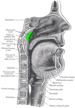
- Pharyngeal tonsil
-
Pharyngeal tonsil 
Location of the adenoid Latin tonsilla pharyngea MeSH Adenoids Adenoids (or pharyngeal tonsil, or nasopharyngeal tonsil) are a mass of lymphoid tissue situated posterior to the nasal cavity, in the roof of the nasopharynx, where the nose blends into the throat.
Normally, in children, they make a soft mound in the roof and posterior wall of the nasopharynx, just above and behind the uvula.
Contents
Histology
Adenoids, unlike other types of tonsils, have pseudostratified columnar ciliated epithelium.[1]
They also differ from the other tonsil types by lacking crypts. The adenoids are often removed along with the tonsils. This can cause a very sore throat for about a week and rather unpleasant breath. Most people's adenoids are not even in use after a person's third year[citation needed], but if they cause problems they must be taken out[citation needed] or they may otherwise shrink.
Pathology
Enlarged adenoids, or adenoid hypertrophy, can become nearly the size of a ping pong ball and completely block airflow through the nasal passages.
Even if enlarged adenoids are not substantial enough to physically block the back of the nose, they can obstruct airflow enough so that breathing through the nose requires an uncomfortable amount of work, and inhalation occurs instead through an open mouth.
Adenoids can also obstruct the nasal airway enough to affect the voice without actually stopping nasal airflow altogether.
Adenoid facies
Enlargement of adenoids, especially in children, causes an atypical appearance of the face, often referred to as adenoid facies.
Features of Adenoid facies include Open mouth/mouth breathing, Long elongated face, prominent incisors, Hypoplastic maxilla Short upper lip, Elevated nostrils, High arched palate,
George Catlin, in his humorous and instructive book Breath of Life, published in 1861, illustrates adenoid faces in many engravings and advocates nose-breathing.[2]
Removal of the adenoids
Surgical removal of the adenoids is a procedure called adenoidectomy.
Adenoids may be removed if they become infected, causing symptoms such as excessive mucus production. Studies have shown that adenoid regrowth occurs in as many as 20% of the cases in which they are removed.
Carried out through the mouth under a general anaesthetic (or less commonly a topical), adenoidectomy involves the adenoids being curetted, cauterised, lasered, or otherwise ablated.
See also
References
- ^ Histology at KUMC lymphoid-lymph06
- ^ Wylie, A, (1927). "Rhinology and laryngology in literature and Folk-Lore". The Journal of Laryngology & Otology 42 (2): 81–87. doi:10.1017/S0022215100029959.
External links
- Roche Lexicon - illustrated navigator, at Elsevier 25420.000-1
- Adenoids: What They Are, How To Recognize Them, What To Do For Them
- Histology at usuhs.mil
- Histology at udel.edu
- /drtbalu otolaryngology online
Head and neck, upper RT: Nose (TA A06.1, TH H3.05.01, GA 10.992) External nose Ala of nose
nasal cartilages (of the septum, Greater alar, Lesser alar, Lateral nasal, Accessory nasal, Vomeronasal)Nasal cavity OpeningsLateral wallNasal concha/meati: Superior nasal concha · Middle nasal concha · Inferior nasal concha · Superior nasal meatus · Middle nasal meatus · Inferior nasal meatus
Sphenoethmoidal recess · Ethmoid bulla · Agger nasi · Ethmoidal infundibulum · Semilunar hiatus · Maxillary hiatusMedial wallParanasal sinuses Naso-pharynx Pharyngeal opening of auditory tube (Salpingopharyngeal fold, Salpingopalatine fold, Torus tubarius) · Pharyngeal tonsil · Pharyngeal recessLymphoid system (TA A13.1–2, TH H3.10, GA 8 and 9) Primary lymphoid organs Secondary lymphoid organs structural: Hilum · Trabeculae · Diaphragmatic surface of spleen · Visceral surface of spleen
Red pulp (Cords of Billroth, Marginal zone)
White pulp (Periarteriolar lymphoid sheaths, Germinal center)
blood flow: Trabecular arteries · Trabecular veinslymph flow: Afferent lymph vessels · Cortical sinuses · Medullary sinuses · Efferent lymph vessels
T cells: High endothelial venules
B cells: Primary follicle/Germinal center · Mantle zone · Marginal zone
layers: Capsule/Trabeculae · Subcapsular sinus · Cortex · Paracortex · Medulla (Medullary cord) · HilumMALT
(process mucosa)M: LMO
anat(h, u, t, a, l)/phys/depv
noco/cong/tumr
proc
Categories:- Lymphatics of the head and neck
Wikimedia Foundation. 2010.
