- Cartilage of the septum
Infobox Anatomy
Name = PAGENAME
Latin = cartilago septi nasi
GraySubject = 223
GrayPage = 992
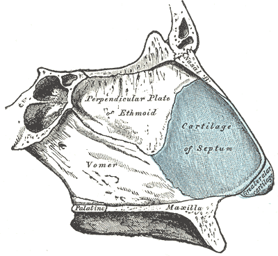
Caption = Bones and cartilages of septum of nose. Right side. (Cartilage of the septum visible as blue structure atright.)

Caption2 = Cartilages of the nose, seen from below. (C. of septum visible in blue at bottom center.)
System =
Precursor =
MeshName =
MeshNumber =
DorlandsPre = c_12
DorlandsSuf = 12217211
The cartilage of the septum (or septal cartilage, or quadrangular cartilage) is somewhat quadrilateral in form, thicker at its margins than at its center, and completes the separation between thenasal cavities in front.Its anterior margin, thickest above, is connected with the
nasal bone s, and is continuous with the anterior margins of thelateral cartilage s; below, it is connected to the medial crura of thegreater alar cartilages by fibrous tissue.Its posterior margin is connected with the perpendicular plate of the
ethmoid ; its inferior margin with the vomer and thepalatine processes of themaxillae .ee also
*
perpendicular plate of ethmoid bone External links
* - "Nasal septum, lateral view"
*
* - "Diagram of skeleton of medial (septal) nasal wall."
* (NormanAnatomyFig|nasalseptumbonescarti)
Wikimedia Foundation. 2010.