- Mantle zone
-
Germinal centre 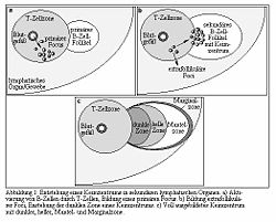
Image labeled in German, but "Mantel-zone" visible near center. The mantle zone (or just mantle) of a lymphatic nodule (or lymphatic follicle) is an outer ring of small lymphocytes surrounding a germinal center.[1]
It is also known as the "corona".[2]
It contains transient lymphocytes.[3]
It is the location of the lymphoma in mantle cell lymphoma.
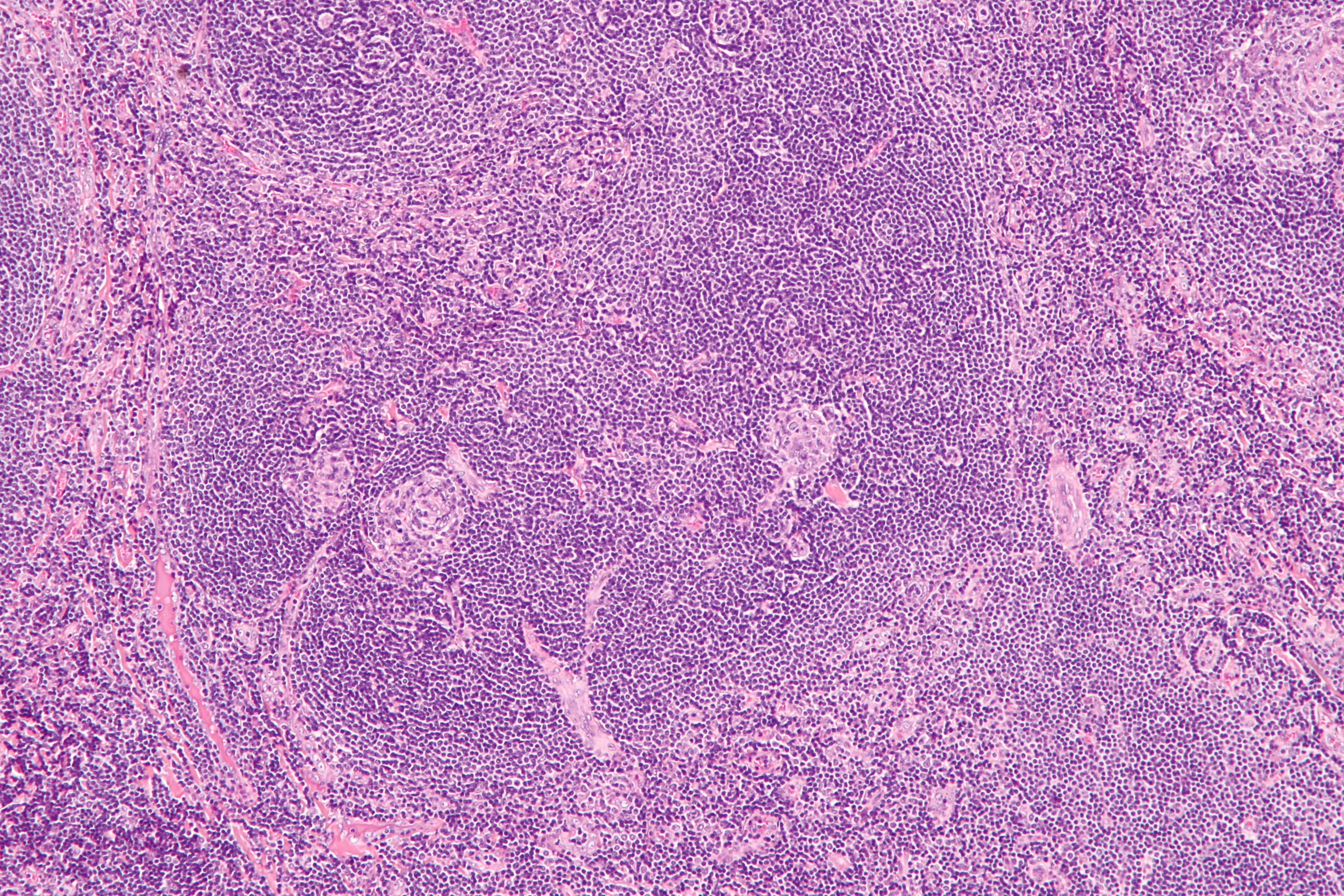
Pathology
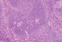 Intermediate magnification micrograph of Castleman disease showing the characteristic expansion of the mantle zone. H&E stain.
Mantle zone expansion may be seen in benign, such as Castleman disease, and malignancy, i.e. Mantle cell lymphoma.
References
External links
- http://erl.pathology.iupui.edu/HISTO/LABE109.HTM
- Histology at BU 07102loa - "Lymphoid Tissues and Organs: lymph node, cortex and medulla"
Lymphoid system (TA A13.1–2, TH H3.10, GA 8 and 9) Primary lymphoid organs Secondary lymphoid organs structural: Hilum · Trabeculae · Diaphragmatic surface of spleen · Visceral surface of spleen
Red pulp (Cords of Billroth, Marginal zone)
White pulp (Periarteriolar lymphoid sheaths, Germinal center)
blood flow: Trabecular arteries · Trabecular veinslymph flow: Afferent lymph vessels · Cortical sinuses · Medullary sinuses · Efferent lymph vessels
T cells: High endothelial venules
B cells: Primary follicle/Germinal center · Mantle zone · Marginal zone
layers: Capsule/Trabeculae · Subcapsular sinus · Cortex · Paracortex · Medulla (Medullary cord) · HilumMALT
(process mucosa)M: LMO
anat(h, u, t, a, l)/phys/depv
noco/cong/tumr
proc
Categories:- Lymphatic system stubs
- Lymphatic organ anatomy

Wikimedia Foundation. 2010.