Trabecular veins

Trabecular veins

Infobox Vein
Name = PAGENAME
Latin =
GraySubject = 278
GrayPage = 1286


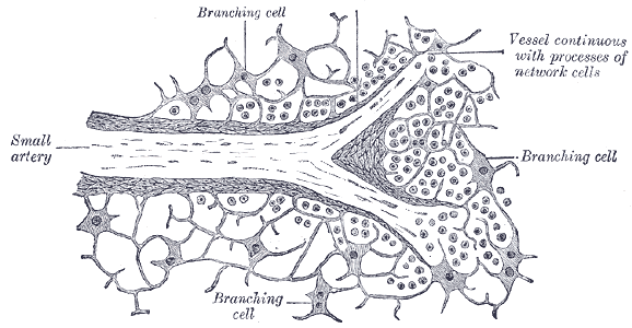
Caption = Section of the spleen, showing the termination of the small bloodvessels.


Caption2 = Transverse section of the spleen, showing the trabecular tissue and the splenic vein and its tributaries.
DrainsFrom = sinus
DrainsTo = Splenic vein
Artery = Trabecular arteries
MeshName =
MeshNumber =
DorlandsPre =
DorlandsSuf =
The trabecular veins are the largest veins inside the spleen. It drains the blood collected in the sinuses of the pulp.

Details

The blood is collected from the interstices of the tissue by the rootlets of the veins, which begin much in the same way as the arteries end.

The connective-tissue corpuscles of the pulp arrange themselves in rows, in such a way as to form an elongated space or sinus.

They become elongated and spindle-shaped, and overlap each other at their extremities, and thus form a sort of endothelial lining of the path or sinus, which is the radicle of a vein.

On the outer surfaces of these cells are seen delicate transverse lines or markings, which are due to minute elastic fibrillæ arranged in a circular manner around the sinus.

Thus the channel obtains an external investment, and gradually becomes converted into a small vein, which after a short course acquires a coat of ordinary connective tissue, lined by a layer of flattened epithelial cells which are continuous with the supporting cells of the pulp.

The smaller veins unite to form larger ones; these do not accompany the arteries, but soon enter the trabecular sheaths of the capsule, and by their junction form six or more branches, which emerge from the hilum, and, uniting, constitute the lienal vein, the largest radicle of the portal vein.

External links

* - "Lymphoid Tissues and Organs: spleen, central artery and trabecular vein"
* [http://www.udel.edu/Biology/Wags/histopage/colorpage/cst/cst.htm Slide at udel.edu]
* (See figure #16)


Wikimedia Foundation. 2010.

Игры ⚽ Нужен реферат?

Look at other dictionaries:

  • trabecular veins — vessels coursing in splenic trabeculae, formed by tributary pulp veins …   Medical dictionary

  • Trabecular arteries — Infobox Artery Name = PAGENAME Latin = GraySubject = 278 GrayPage = 1285 Caption = Transverse section of the human spleen, showing the distribution of the splenic artery and its branches. Caption2 = Section of the spleen, showing the termination… …   Wikipedia

  • Trabecular meshwork — Enlarged general view of the iridial angle. (When enlarged, visible with older label of trabecular tissue ) Latin reticulum trabeculare sclerae Gray s …   Wikipedia

  • Lymphoid system — Lymphatic drainage redirects here. For the massage technique, see Manual lymphatic drainage. Lymphoid System An image displaying the lymphoid system. Latin …   Wikipedia

  • White pulp — Transverse section of a portion of the spleen. (Lymphatic nodule labeled at center right.) …   Wikipedia

  • Bone marrow — Simplified illustration of cells in bone marrow. Latin medulla ossium Code TA …   Wikipedia

  • Haematopoiesis — Diagram showing the development of different blood cells from haematopoietic stem cell to mature cells Haematopoiesis (from Ancient Greek: αἷμα, blood ; ποιεῖν to make ) (or hematopoiesis in the United States; sometimes also haemopoiesis or… …   Wikipedia

  • Immune system — A scanning electron microscope image of a single neutrophil (yellow), engulfing anthrax bacteria (orange). An immune system is a system of biological structures and processes within an organism that protects against disease by identifying and… …   Wikipedia

  • Pharyngeal tonsil — Location of the adenoid Latin tonsilla pharyngea MeSH …   Wikipedia

  • Cords of Billroth — The Cords of Billroth (also known as splenic cords or red pulp cords) are found in the red pulp of the spleen between the sinusoids, consisting of fibrils and connective tissue cells with a large population of monocytes and macrophages. These… …   Wikipedia

Share the article and excerpts

Direct link
Do a right-click on the link above
and select “Copy Link”