- Meniscus (anatomy)
-
Meniscus (anatomy) 
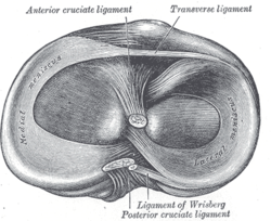
Head of right tibia seen from above, showing menisci and attachments of ligaments 
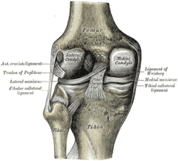
Left knee-joint from behind, showing interior ligaments In anatomy, a meniscus (from Greek μηνίσκος meniskos, "crescent"[1]) is a crescent-shaped fibrocartilaginous structure that, in contrast to articular disks, only partly divides a joint cavity.[2] In humans it is present in the knee, acromioclavicular, sternoclavicular, and temporomandibular joints;[3] in other organisms they may be present in other joints (e.g. between the forearm bones of birds). A small meniscus also occurs in the radio-carpal joint.
It usually refers to either of two specific parts of cartilage of the knee: The lateral and medial menisci. Both are cartilaginous tissues that provide structural integrity to the knee when it undergoes tension and torsion. The menisci are also known as 'semi-lunar' cartilages — referring to their half-moon "C" shape — a term which has been largely dropped by the medical profession, but which led to the menisci being called knee 'cartilages' by the lay public.
Contents
Anatomy
The menisci of the knee joint are two pads of cartilaginous tissue which serve to disperse friction in the knee joint between the lower leg (tibia) and the thigh (femur). They are shaped concave on the top and flat on the bottom, articulating with the tibia. They are attached to the small depressions (fossae) between the condyles of the tibia (intercondyloid fossa), and towards the center they are unattached and their shape narrows to a thin shelf.[4]
Function
The menisci act to disperse the weight of the body and reduce friction during movement. Since the condyles of the femur and tibia meet at one point (which changes during flexion and extension), the menisci spread the load of the body's weight.[5] This differs from sesamoid bones, which are made of osseous tissue and whose function primarily is to protect the nearby tendon and to increase its mechanical effect.
Injury
Main article: Tear of meniscusIn sports and orthopedics, people will sometimes speak of "torn cartilage" and actually be referring to an injury to one of the menisci.
The Unhappy Triad is a set of commonly co-occurring knee injuries which includes injury to the medial meniscus.
Two surgeries are most common of the meniscus. Depending on location, age, and doctor's preference, people usually either repair it or remove a part or all of it (meniscectomy). Each has its advantages and disadvantages.
See also
Notes
- ^ μηνίσκος, "small moon", is diminutive of μήνη, "moon", from the root ma-, "measure", which reflects the fact the time was measured according to the phases of the moon. The word was also used for curved things in general, such as a necklace or a line of battle. (Lexicon of Orthopaedic Etymology, p 199)
- ^ Platzer (2004), p 208
- ^ Meniscus, Stedman's (27th ed.)
- ^ Gray's (1918), 7b
- ^ Cluett, Meniscus Tear — Torn Cartilage
References
- "Meniscus". Stedman's Medical Dictionary, 27th edition. eMedicine - Lippincott Williams & Wilkins. 2003. http://www.webcitation.org/5VlKiXRhC. Retrieved 2008-02-20.
- Cluett, Jonathan (February 10, 2008). "Meniscus Tear — Torn Cartilage". About.com. http://www.webcitation.org/5VlLgNp5d. Retrieved 2008-02-20.
- Diab, Mohammad (1999). Lexicon of Orthopaedic Etymology. Taylor & Francis. ISBN 9057025973. http://books.google.com/books?id=fstFQVnw8-wC&pg=PA200.
- Gray, Henry (1918). "7b. The Knee-joint". Gray's Anatomy of the Human Body. http://www.webcitation.org/5VlLVEO4e. Retrieved 2008-02-20.[broken citation]
- Platzer, Werner (2004). Color Atlas of Human Anatomy, Vol. 1: Locomotor System (5th ed.). Thieme. ISBN 3-13-533305-1.
External links
- Athletic advisor
- Arthroscopy. com about torn menisci
- Orthogate on meniscus injuries
- Orthogate on Meniscal Surgery
Joints (TA A03.0, TH H3.02, GA 3.284) Types fibrous: Gomphosis · Suture · Syndesmosis · Interosseous membrane
cartilaginous: Synchondrosis · Symphysis
synovial: Plane joint · 1° (Hinge joint, Pivot joint) · 2° (Condyloid joint, Saddle joint) · 3° (Ball and socket joint)
by range of motion: Synarthrosis · Amphiarthrosis · DiarthrosisTerminology Motions general: Flexion/Extension · Adduction/Abduction · Internal rotation/External rotation · Elevation/Depression
specialized/upper limbs: Protraction/Retraction · Supination/Pronation
specialized/lower limbs: Plantarflexion/Dorsiflexion · Eversion/InversionComponents capsular: Articular capsule (Synovial membrane, Fibrous membrane) · Synovial fluid · Synovial bursa · Articular disk/Meniscus
extracapsular: Ligament · EnthesisM: JNT
anat(h/c, u, t, l)/phys
noco(arth/defr/back/soft)/cong, sysi/epon, injr
proc, drug(M01C, M4)
Joints and ligaments of lower limbs (TA A03.6, GA 3.333) Coxal/hip femoral (iliofemoral, pubofemoral, ischiofemoral) · head of femur · transverse acetabular · acetabular labrum · capsule · zona orbicularisKnee-joint TibiofemoralCapsule · Anterior meniscofemoral ligament · Posterior meniscofemoral ligament
extracapsular: popliteal (oblique, arcuate) · collateral (medial/tibial, fibular/lateral)
intracapsular: cruciate (anterior, posterior) · menisci (medial, lateral) · transversePatellofemoralTibiofibular Superior tibiofibularInferior tibiofibularJoints of foot medial: medial of talocrural joint/deltoid (anterior tibiotalar, posterior tibiotalar, tibiocalcaneal, tibionavicular)
lateral: lateral collateral of ankle joint (anterior talofibular, posterior talofibular, calcaneofibular)Distal intertarsalOtherM: JNT
anat(h/c, u, t, l)/phys
noco(arth/defr/back/soft)/cong, sysi/epon, injr
proc, drug(M01C, M4)
Categories:- Joints
Wikimedia Foundation. 2010.

