- Iliofemoral ligament
Infobox Ligament
Name = Iliofemoral ligament
Latin = ligamentum iliofemorale
GraySubject = 92
GrayPage = 335

Caption = Right hip-joint from the front. (Iliofemoral ligament visible at center.)
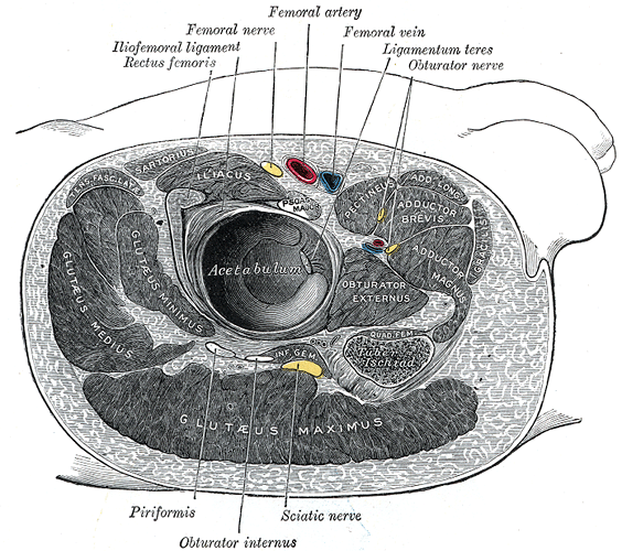
Caption2 = Structures surrounding right hip-joint. (Iliofemoral ligament labeled at upper left.)
From = ilium (anterior inferior iliac spine )
To =femur (intertrochanteric line )
MeshName =
MeshNumber =
DorlandsPre = l_09
DorlandsSuf = 12492337
The iliofemoral ligament is aligament of thehip joint which extends from the ilium to thefemur in front of the joint. It is also referred to as the Y-ligament (see below) or the ligament of Bigelow, and any combinations of these names.With a tensile strength exceeding 350 N, the iliofemoral ligament is not only stronger than the two other ligaments of the hip joint, the ischiofemoral and the pubofemoral, but also the strongest ligament in the human body and as such is an important constraint to the hip joint."Thieme Atlas of Anatomy" (2006), p 380]
Structure
Arising from the
anterior inferior iliac spine and the rim of theacetabulum , the iliofemoral ligament spreads obliquely downwards and lateralwards to theintertrochanteric line on the anterior side of the femoral head. It is divided into two parts or bands who act differently: The transversal part above, is strong and runs parallel to the axis of the femoral neck. The descending part below, is weaker and runs parallel to the femoral shaft. As the lateral portion is twisted like a screw, the two parts together take the form an inverted Y.Platzer (2004), p 200]it is intimately connected with the
joint capsule , and serves to strengthen the joint by resistinghyperextension . Its upper band is sometimes named the iliotrochanteric ligament. Between the two bands is a thinner part of the capsule. In some cases there is no division, and the ligament spreads out into a flat triangular band which is attached to the whole length of the intertrochanteric line.Function
In a standing posture, when the
pelvis is tilted posteriorly, the ligament is twisted and tense, which prevents the trunk from falling backwards and the posture is maintained without the need for muscular activity. In this position the ligament also keeps the femoral head pressed into the acetabulum.As the thighs flexes, the tension in the ligament is reduced and the amount of possible rotations in the hip joint is increased, which permits the pelvis to tilt backwards into its sitting angle. Lateral rotation and adduction in the hip joint is controlled by the strong transversal part, while the descending part limits medial rotation.
Turnout used in the classical
ballet style requires a great deal of flexibility in this ligament.Additional images
Notes
References
* cite book
title = Color Atlas of Human Anatomy, Vol 1: Locomotor system
first = Werner | last = Platzer
edition = 5th | publisher = Thieme
isbn = 3-13-533305-1 | year = 2004 (ISBN for the Americas 1-58890-159-9.)
* cite book
title = Thieme Atlas of Anatomy
publisher = Thieme
isbn = 3-13-1420511-2 | year = 2006 (ISBN for the Americas 1-58890-419-9)External links
* (NormanAnatomyFig|hipjointanterior)
*
Wikimedia Foundation. 2010.