- Intercuneiform articulations
-
Intercuneiform and cuneocuboid articulations 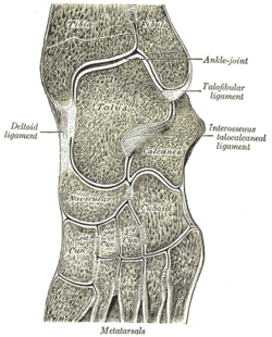
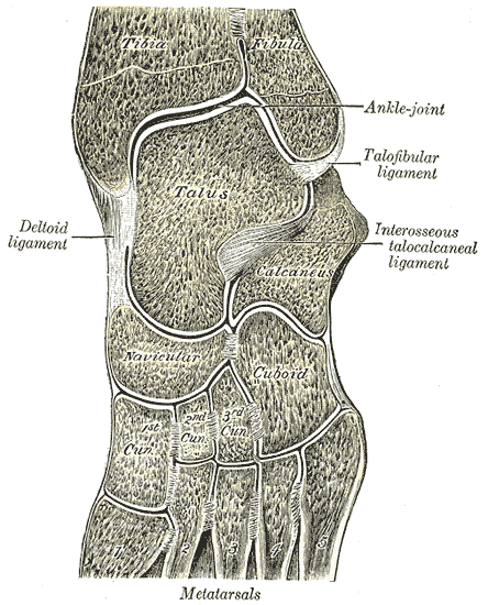
Oblique section of left intertarsal and tarsometatarsal articulations, showing the synovial cavities. Latin articulationes intercuneiformes Gray's subject #96 357 The intercuneiform articulations are articulations among the cuneiform bones.
The term "cuneocuboid articulation" is sometimes used to describe the joint between the cuboid and lateral cuneiform, but this term isn't recognized by Terminologia Anatomica.
Contents
Ligaments
The three cuneiform bones and the cuboid are connected together by dorsal, plantar, and interosseous ligaments.
The Dorsal Ligaments
The dorsal ligaments consist of three transverse bands: one connects the first with the second cuneiform, another the second with the third cuneiform, and another the third cuneiform with the cuboid.
The Plantar Ligaments
The plantar ligaments have a similar arrangement to the dorsal, and are strengthened by slips from the tendon of the Tibialis posterior.
The Interosseous Ligaments
The interosseous ligaments consist of strong transverse fibers which pass between the rough non-articular portions of the adjacent surfaces of the bones.
Synovial Membrane
The synovial membrane of these joints is part of the great tarsal synovial membrane.
Movements
The movements permitted between these bones are limited to a slight gliding upon each other.
References
This article was originally based on an entry from a public domain edition of Gray's Anatomy. As such, some of the information contained within it may be outdated.
Joints and ligaments of lower limbs (TA A03.6, GA 3.333) Coxal/hip femoral (iliofemoral, pubofemoral, ischiofemoral) · head of femur · transverse acetabular · acetabular labrum · capsule · zona orbicularisKnee-joint TibiofemoralCapsule · Anterior meniscofemoral ligament · Posterior meniscofemoral ligament
extracapsular: popliteal (oblique, arcuate) · collateral (medial/tibial, fibular/lateral)
intracapsular: cruciate (anterior, posterior) · menisci (medial, lateral) · transversePatellofemoralTibiofibular Superior tibiofibularInferior tibiofibularJoints of foot medial: medial of talocrural joint/deltoid (anterior tibiotalar, posterior tibiotalar, tibiocalcaneal, tibionavicular)
lateral: lateral collateral of ankle joint (anterior talofibular, posterior talofibular, calcaneofibular)Distal intertarsalIntercuneiformOtherM: JNT
anat(h/c, u, t, l)/phys
noco(arth/defr/back/soft)/cong, sysi/epon, injr
proc, drug(M01C, M4)
Categories:- Lower limb anatomy
- Joints
- Musculoskeletal system stubs
Wikimedia Foundation. 2010.