- Tear of meniscus
-
Tear of meniscus Classification and external resources 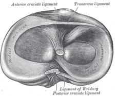
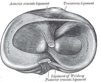
Head of right tibia seen from above, showing menisci and attachments of ligamentsICD-10 Current injury S83.2
Old tear M23.2ICD-9 836.0-836.2 In sports and orthopedics, a tear of a meniscus is a rupturing of one or more of the fibrocartilage strips in the knee called menisci. When doctors and patients refer to "torn cartilage" in the knee, they actually may be referring to an injury to a meniscus at the top of one of the tibiae. Menisci can be torn during innocuous activities such as walking or squatting. They can also be torn by traumatic force encountered in sports or other forms of physical exertion. The traumatic action is most often a twisting movement at the knee while the leg is bent. In older adults, the meniscus can be damaged following prolonged 'wear and tear' called a degenerative tear.
Tears can lead to pain and/or swelling of the knee joint. Especially acute injuries (typically in younger, more active patients) can lead to displaced tears which can cause mechanical symptoms such as clicking, catching, or locking during motion of the knee joint.[1] The joint will be in pain when in use, but when there is no load, the pain goes away.
A tear of the medial meniscus can occur as part of the unhappy triad, together with a tear of the anterior cruciate ligament and medial collateral ligament.
Contents
Anatomy
The menisci are C-shaped wedges of fibrocartilage located between the tibial plateau and femoral condyles. The menisci contain 70% type I collagen.[2] The larger semilunar medial meniscus is attached more firmly than the loosely fixed, more circular lateral meniscus. The anterior and posterior horns of both menisci are secured to the tibial plateaus. Anteriorly, the transverse ligament connects the 2 menisci; posteriorly, the meniscofemoral ligament helps stabilize the posterior horn of the lateral meniscus to the femoral condyle. The coronary ligaments connect the peripheral meniscal rim loosely to the tibia. Although the lateral collateral ligament (LCL) passes in close proximity, the lateral meniscus has no attachment to this structure.[2]
The joint capsule attaches to the entire periphery of each meniscus but adheres more firmly to the medial meniscus. An interruption in the attachment of the joint capsule to the lateral meniscus, forming the popliteal hiatus, allows the popliteus tendon to pass through to its femoral attachment site. Contraction by the popliteus during knee flexion pulls the lateral meniscus posteriorly, avoiding entrapment within the joint space. The medial meniscus does not have a direct muscular connection. The medial meniscus may shift a few millimeters, while the less stable lateral meniscus may move at least 1 cm.
In 1978, Shrive et al. reported that the collagen fibers of the menisci are oriented in a circumferential pattern.[2] When a compressive force is applied in the knee joint, a tensile force is transmitted to the menisci. The femur attempts to spread the menisci anteroposteriorly in extension and mediolaterally in flexion. Shrive et al. further studied the effects of a radial cut in the peripheral rim of the menisci during loading. In joints with intact menisci, the force was applied through the menisci and articular cartilage; however, a lesion in the peripheral rim disrupted the normal mechanics of the menisci and allowed it to spread when a load was applied. The load now was distributed directly to the articular cartilage. In light of these findings, it is essential to preserve the peripheral rim during partial meniscectomy to avoid irreversible disruption of the structure's hoop tension capability.[2]
Symptoms and signs
The patient's chief complaints are usually knee pain and swelling. These are worse when the knee bears more weight (for example, when running). Another typical complaint is joint locking, when the patient is unable to fully straighten the leg. This can be accompanied by a clicking feeling. Sometimes, a meniscal tear also causes a sensation that the knee gives way.
The patient can sometimes remember a specific activity during which the injury was sustained. A tear of the meniscus commonly follows a trauma which involves rotation of the knee while it was slightly bent. These maneuvers also excite the pain after the injury; for example, getting out of a car is often reported as painful.
After noting symptoms, a physician can perform clinical tests to determine if the pain is caused by compression and impingement of a torn meniscus. The knee is examined for swelling. In meniscal tears, pressing on the joint line on the affected side typically produces tenderness. The McMurray test involves pressing on the joint line while stressing the meniscus (using flexion-extension movements and varus or valgus stress). Similar tests are the Steinmann test (with the patient sitting) and the Appley test (a grinding maneuver while the patient lies prone and the knee is bent 90°). Bending the knee (into hyperflexion if tolerable), and especially squatting, is typically a painful maneuver if the meniscus is torn. The range of motion of the joint is often restricted.
The Cooper's sign is present in over 92% of tears. It is a subjective symptom of pain in the affected knee when turning over in bed at night. Osteoarthritic pain is present with weightbearing, but the meniscal tear causes pain with a twisting motion of the knee as the meniscal fragment gets pinched, and the capsular attachment gets stretched causing the complaint of pain.
Also see: bounce home test, Thessally's test, Wilson test (internal/external rotation), and Bohler's test.
Classification of Meniscal tear
A Meniscus tear can be classified in various ways – by anatomic location, by proximity to blood supply, etc. Various tear patterns and configurations have been described.[3] These include:
  • Radial tears
  • Flap or Parrot-beak tears
  • Peripheral, longitudinal tears
  • Bucket-handle tears
  • Horizontal cleavage tears
  • Complex, degenerative tears
These tears can then further classified by their proximity to meniscus blood supply, namely whether they are located in the “red-red,” “red-white,” or “white-white” zones.
The functional importance of these classifications, however, is to ultimately determine whether a meniscus is REPAIRABLE. The repairability of a meniscus is dependent upon a number of factors. These include:
  • Age/strength
  • Activity Level
  • Tear Pattern
  • Chronicity of the tear
  • Associated Injuries (Anterior Cruciate Ligament Injury)
  • Healing Potential
Causes
There are two menisci in your knee. They sit between the thigh bone femur and shin bone tibia. While the ends of the thigh bone and shin bone have a thin covering of soft hyaline cartilage, the menisci are made of fibrocartilage tough cartilage and conform to the surfaces of the bones upon which they rest. One meniscus rests on the medial tibial plateau; this is the medial meniscus. The other meniscus rests on the lateral tibial plateau; this is the lateral meniscus.[4]
These menisci act to distribute body weight across the knee joint. Without the menisci, the weight of the body would be unevenly applied to the bones in the legs (the femur and tibia). This uneven weight distribution would cause the development of abnormal excessive forces leading to early damage of the knee joint. The menisci also contribute to the stability of the joint.
The menisci are nourished by small blood vessels, but the menisci also have a large area in the center of that has no direct blood supply (avascular). This presents a problem when there is an injury to the meniscus as the avascular areas tend not to heal. Without the essential nutrients supplied by blood vessels, healing cannot take place.[4]
The two most common causes of a meniscus tear are traumatic injury (often seen in athletes) and degenerative processes (seen in elderly patients who have more brittle cartilage).[4] Meniscus tears can occur in all age groups. Traumatic tears are most common in active people from age 10-45.[4] Traumatic meniscal tears are usually radial or vertical in the meniscus and are more likely to produce a moveable fragment that can catch in the knee and therefore require surgical treatment.
The most common mechanism of a traumatic meniscus tear occurs when the knee joint is bent and the knee is then twisted. It is not uncommon for the meniscus tear to occur along with injuries to the anterior cruciate ligament ACL and the medial collateral ligament MCL — these three problems occurring together are known as the "unhappy triad," which is seen in sports such as football when the player is hit on the outside of the knee. Individuals who experience a meniscus tear usually experience pain and swelling as their primary symptoms. Another common complaint is joint locking, or the inability to completely straighten the joint. This is due to a piece of the torn cartilage preventing the normal functioning of the knee joint.
Degenerative tears are most common in people from age 40 upward. Degenerative meniscal tears are thought to occur as part of the aging process when the collagen fibers within the meniscus start to break down and lend less support to the structure of the menicus. Degenerative tears are usually horizontal in the meniscus, producing both an upper and lower segment of meniscus. These segments usually don't move out of place and therefore are less likely to produce mechanical symptoms of catching or locking.[4]
Degenerative meniscal tears are also more common in smokers.
A torn meniscus can prevent normal pain-free motion of the knee and therefore can interfere with the patient's ability to climb stairs or get in and out of chairs and cars.
Pathophysiology
The medial and lateral menisci are shock absorbers and force distributors located between the femur and the tibia. Consequently, menisci can tear due to traumatic injury or degenerative wear (e.g., in knee joint arthritis), and can compromise force distribution across the knee joint, increasing force concentration on the cartilage and other joint structures.
Damage to the meniscus is due to rotational forces directed to a flexed knee (as may occur with twisting sports) is the usual underlying mechanism of injury. A valgus force applied to a flexed knee with the foot planted and the femur rotated externally can result in a lateral meniscus tear. A varus force applied to the flexed knee when the foot is planted and the femur rotated internally can result in a tear of the medial meniscus.
Tears produce rough surfaces inside the knee, which cause catching, locking, buckling, pain, or a combination of these symptoms. Abnormal loading patterns and rough surfaces inside the knee, especially when coupled with return to sports, significantly increase the risk of developing arthritis if not already present.
Diagnosis
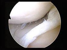
X-ray images (normally during weightbearing) can be obtained to rule out other conditions or to see if the patient also has osteoarthritis. The menisci themselves cannot be visualised with plain radiographs. If the diagnosis is not clear from the history and examination, the menisci can be imaged with magnetic resonance imaging (an MRI scan). This technique has replaced previous arthrography, which involved injecting contrast medium into the joint space. In straightforward cases, knee arthroscopy allows quick diagnosis and simultaneous treatment. Recent clinical data shows that MRI and clinical testing are comparable in sensitivity and specificity when looking for a meniscal tear.
Treatment
 Tear of medial meniscus
Conservative
The treatment course is dependent on the needs and status of the patient. A conservative course of treatment involving just physical therapy is possible. The patient will probably have to take a small break from his or her normal activities, allowing the knee to heal. Exercises can strengthen the muscles around the knee, especially the quadriceps. Stronger and bigger muscles will protect the meniscus cartilage by absorbing a part of the weight. The patient may be given paracetamol or anti-inflammatory medications.
Surgery
If this does not resolve the symptoms or in cases of a locked knee, then surgical intervention may be required. Depending on the location of the tear, a repair may be possible. In the outer third of the meniscus, an adequate blood supply exists and a repair will likely heal.[1] Usually younger patients are more resilient and respond well to this treatment, while older, more sedentary patients do not have a favorable outcome after a repair.[5]
The meniscus has fewer vessels and blood flow towards the unattached, thin interior edge. In the majority of cases, the tear is far away from the meniscus' blood supply, and a repair is unlikely to heal. In these cases arthroscopic surgery allows for a partial meniscectomy, removing the torn tissue and allowing the knee to function with some of the meniscus missing. In situations where the meniscus is damaged beyond repair or partial removal, a total meniscectomy is performed. This option is avoided at all costs as total meniscectomy leads to an increased risk of osteoarthritis (with loss of cartilage) and eventual total knee replacement. In some cases, a meniscus replacement is done to prevent this.
Transplants of full meniscus are accomplished successfully regularly, although it is still somewhat of a rare procedure and many questions surrounding its use remain.[6][5]
Post-surgical rehabilitation
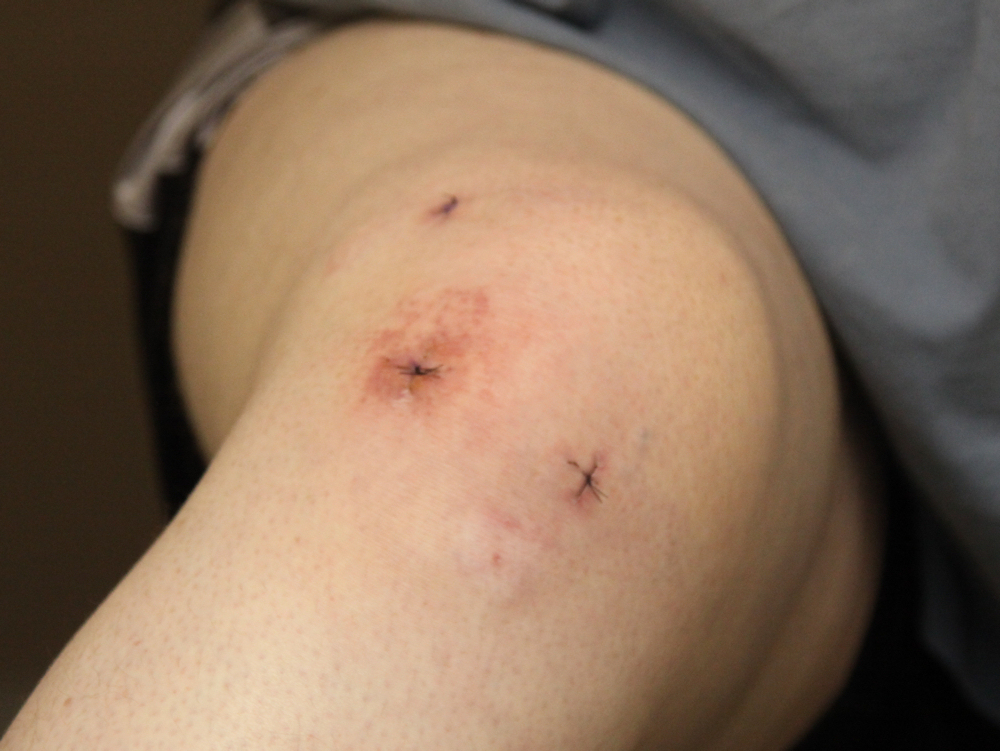
 Typical locations of arthroscopic surgery incisions in a knee joint following surgery for a tear in the meniscus.
After a successful surgery for treating the destroyed part of the meniscus patients must follow a rehabilitation program to have the best result. The rehabilitation following a meniscus surgery depends on whether the entire meniscus was removed or repaired. Although not clinically proven some people report better recovery after a period of oral intake of glucosamine-sulphate supplement.[dubious ]
If the destroyed part of the meniscus was removed, patients can usually start walking using a crutch a day or two after surgery. Although each case is different, patients return to their normal activities on average after a few weeks (2 or 3). Still, completely normal walk will resume gradually and it's not unusual to take 2–3 months for the recovery to reach a level where a patient will walk totally smoothly. Many meniscectomy patients don't ever feel a 100% functional recovery, but even years after the procedure they sometimes feel tugging or tension in a part of their knee. There is little medical follow-up after meniscectomy and official medical documentation tends to ignore the imperfections and side-effects of this procedure.
If the meniscus was repaired the rehabilitation program that follows is a lot more intensive. After the surgery a hinged knee brace is sometimes placed on the patient. This brace allows controlled movement of the knee. The patient is encouraged to walk using crutches from the first day, and most of the times can put partial weight on the knee.
The time course varies with each patient. Starting from the second month the patient maybe able to walk freely and can also do various "low-impact" exercises (static bicycle, swimming, etc.), but should expect the knee to feel stiff and sore. If the rehabilitation was done properly the patient can gradually return back to "high-impact" activities (like running). However, "heavier activities", like running, skiing, basketball etc., generally any activities where knees bear sudden changes of the direction of movement can lead to repeated injuries. When planning sport activities it makes sense to consult a physical therapist and check how much impact the sport will have on the knee.
Epidemiology
In 2008 the U.S Department of Health and Human Services reported a combined total of 2,295 discharges for the principal diagnosis of tear of lateral cartilage/meniscus (836.0), tear of medial cartilage/meniscus (836.1), and tear of cartilage/meniscus (836.2). Females had a total of 53.49% discharges while males had 45.72%. Individuals between the ages of 45-68 year had an average of 31.73% discharges followed by age group 65-84 with 28.82%. The average length of stay for a patient diagnosed with torn menisci was 2.7 days for males and 3.7 days for females. There was a report of 6,941 hospital discharges for knee repair. Individuals between 18-44 years of age were among the highest with 37.37% total of discharges followed by the age group 45-64 with a percentage of 36.34%. Males had a slightly higher number of discharges (50.78%) than females (48.66%). The average length of stay for both male and female patients in a hospital setting was 3.1.[7]
See also
References
- ^ a b Meniscus Injuries - eMedicine.
- ^ a b c d "Anatomy of Meniscus" 1994. May. 2011. Anatomy, Meniscal Tear.
- ^ Classification of meniscal tear, sportsmd.
- ^ a b c d e ”Causes of meniscus tear”. 2004. Web. May. 2011 Causes of meniscus tear.
- ^ a b Sohn DH, Toth AP (April 2008). "Meniscus transplantation: current concepts". J Knee Surg 21 (2): 163–72. PMID 18500070.
- ^ Matava MJ (February 2007). "Meniscal allograft transplantation: a systematic review". Clin. Orthop. Relat. Res. 455: 142–57. doi:10.1097/BLO.0b013e318030c24e. PMID 17279042. http://meta.wkhealth.com/pt/pt-core/template-journal/lwwgateway/media/landingpage.htm?doi=10.1097/BLO.0b013e318030c24e.
- ^ “Epidemiology of meniscus”. 2006. Web. May. 2011 [http://www.injuryjournal.com/article/S0020-1383%2808%2900298-2/abstract.
Musculoskeletal disorders: Acquired musculoskeletal deformities (M20–M25, M95, 734–738) Upper limb Lower limb foot deformity (Bunion/hallux valgus, Hallux varus, Hallux rigidus, Hammer toe, Foot drop, Flat feet, Club foot)Genu recurvatumHead General terms M: JNT
anat(h/c, u, t, l)/phys
noco(arth/defr/back/soft)/cong, sysi/epon, injr
proc, drug(M01C, M4)
Injury: Dislocations/subluxations, sprains and strains (Sx3 where x=0 to 9, 830–848) Joints and
ligamentsHead and neckShoulder and upper armMuscles and
tendonsShoulder and upper armM: JNT
anat(h/c, u, t, l)/phys
noco(arth/defr/back/soft)/cong, sysi/epon, injr
proc, drug(M01C, M4)
Categories:- Dislocations, sprains and strains
- Injuries of knee and lower leg

Wikimedia Foundation. 2010.