- Rotator cuff tear
Infobox_Disease
Name = PAGENAME
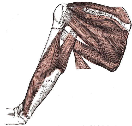
Caption = Muscles on the dorsum of the scapula, and the Triceps brachii.
DiseasesDB = 32230
ICD10 = ICD10|M|75|1|m|70, ICD10|S|46|0|s|40
ICD9 = ICD9|726.1 ICD9|727.61, ICD9|840.4
ICDO =
OMIM =
MedlinePlus =
eMedicineSubj = radio
eMedicineTopic = 894
eMedicine_mult = eMedicine2|pmr|125 eMedicine2|radio|889 eMedicine2|sports|115
MeshID =Rotator cuff tears are tears of one, or more, of the four
tendon s of therotator cuff muscles.Rotator cuff tears are among the most common conditions affecting the shoulder.cite journal |author=Williams GR, Rockwood CA, Bigliani LU, Iannotti JP, Stanwood W |title=Rotator cuff tears: why do we repair them? |journal=J Bone Joint Surg Am |volume=86-A |issue=12 |pages=2764–76 |year=2004 |pmid=15590865 |doi= |url=http://www.ejbjs.org/cgi/pmidlookup?view=long&pmid=15590865]
The tendons of the rotator cuff, not the muscles, are most commonly torn. Of the four tendons, the supraspinatus is most frequently torn; the tear usually occurs at its point of insertion onto the humeral head at the greater tuberosity.cite web |url=http://orthoinfo.aaos.org/topic.cfm?topic=A00406&return_link=0 |title=Your Orthopaedic Connection: Rotator Cuff Tears and Treatment Options |format= |work= |accessdate=]
Anatomy
The rotator cuff muscles, a group of four muscles that surround the shoulder, are the:
supraspinatus ,infraspinatus ,teres minor andsubscapularis . The four rotator cuff muscle tendons combine to form a broad, conjoined tendon, called the rotator cuff tendon, and insert onto the bone of the humeral head in the shoulder. The humeral head is the ball side of the “ball and socket ” shoulder joint; the socket is called theglenoid fossa .Presentation
Many rotator cuff tears cause no pain nor produce any symptoms, tears are known to have an increasing incidence with increasing age. The most frequent cause of rotator cuff damage is age related degeneration and less frequently by
sports injuries or trauma. Partial and full thickness tears have been found on post mortem studies and on MRI studies, in people who do not have a history of shoulder pain or symptoms.Classification
Tears of the rotator cuff tendon are described as partial thickness tears, full thickness tears and full thickness tears with complete detachment of the tendons from bone.
* Partial thickness tears often appear as fraying of an intact tendon.
* Full thickness tears are through-and-through tears. These can be small pin-point tears or larger button hole tears or tears involving the majority of the tendon where the tendon still remains substantially attached to the humeral head and thus maintains function.
* Full thickness tears may may also involve complete detachment of the tendon(s) from the humeral head and may result in impaired shoulder motion and function may be significantly affected.
Shoulder pain is variable and does not always correspond to the size of the tear.
Prognosis
While people with rotator cuff tears may not have any noticeable symptoms, studies have shown that over time 40% will have enlargement of the tear over a 5-year period. Of those whose tears enlarge, 20% have no symptoms while 80% eventually develop symptoms. [cite journal |author=Tempelhof S, Rupp S, Seil R |title=Age-related prevalence of rotator cuff tears in asymptomatic shoulders |journal=J Shoulder Elbow Surg |volume=8 |issue=4 |pages=296–9 |year=1999 |pmid=10471998 |doi= |url=]
Diagnosis
The in-person clinical evaluation has two parts: the history and the physical examination.The history records the patient’s stated symptoms. The physical examination documents physical signs.
For rotator cuff tears the history is variable and may include a discreet episode of trauma or no trauma at all. The pain may have started suddenly or may have come on gradually. The pain may be constant, intermittent, or only activity related. The pain may be mild to severe and weakness may or may not be noted.
ymptoms associated with rotator cuff tears:
The most reliable symptom for determining a rotator cuff tear is probably the least common and is found when there is a complete rupture with detachment of the rotator cuff leading to the complaint of complete loss of function, such as, loss of the ability to actively move the arm away from the side of the body (loss of abduction). Fortunately this finding is rare and when tears are symptomatic, most tears present as pain with limitation of function, a non-specific complaint that cannot distinguish between tendinitis, bursitis or arthritis. The clinical picture of a completely detached tear is more clear-cut, while the more common shoulder problems greatly overlap in their clinical presentation.
Pain in the anterolateral aspect of the shoulder can be due to many causes, [cite journal |author=McFarland EG, Selhi HS, Keyurapan E |title=Clinical evaluation of impingement: what to do and what works |journal=J Bone Joint Surg Am |volume=88 |issue=2 |pages=432–41 |year=2006 |pmid=16475277 |doi= |url=http://www.ejbjs.org/cgi/pmidlookup?view=long&pmid=16475277] symptoms may reflect pathology outside of the shoulder which cause referred pain to the shoulder from sites such as the neck, heart or gut.
Patient history will often include pain or ache over the front and outer aspect of the shoulder, pain aggravated by leaning on the elbow and pushing upwards on the shoulder (such as leaning on the armrest of a reclining chair), intolerance to overhead activity, pain at night when laying directly on the affected shoulder, pain when reaching forward (e.g. unable to lift a gallon of milk from the refrigerator). Weakness may be reported, but is often masked by pain and is usually found only through examination. With longer standing pain, the shoulder is favored and gradually loss of motion and weakness may develop which, due to pain and guarding are often missed by the patient and are only brought out during the examination.
Primary shoulder problems may cause pain over the deltoid muscle that is made worse by abduction against resistance, called the impingement sign. Impingement reflects pain arising from the rotator cuff but cannot distinguish between inflammation, strain, or tear. Patients may report their experience with the impingement sign when they report that they are unable to reach upwards to brush their hair or to reach in front to lift a can of beans up from an overhead shelf.
igns associated with rotator cuff tears:
A paper in the medical literature in 2006 cite journal |author=Park HB, Yokota A, Gill HS, El Rassi G, McFarland EG |title=Diagnostic accuracy of clinical tests for the different degrees of subacromial impingement syndrome |journal=J Bone Joint Surg Am |volume=87 |issue=7 |pages=1446–55 |year=2005 |pmid=15995110 |doi=10.2106/JBJS.D.02335 |url=http://www.ejbjs.org/cgi/pmidlookup?view=long&pmid=15995110] , evaluated eight well known physical examination tests to determine their diagnostic values to help distinguish between bursitis, partial-thickness rotator cuff tears, and full-thickness rotator cuff tears. The study concluded that, "the best test" was a combination of tests. For the diagnosis of impingement disease the best combination of tests were a positive: Hawkins-Kennedy impingement sign, a positive painful arc sign, and weakness in external rotation with the arm at the side. To diagnose a full-thickness rotator cuff tear, the best combination of tests, when all three are positive: were the painful arc, the drop-arm sign, and weakness in external rotation .
Diagnostic tests
The correct use of diagnostic tests is an important component of effective medical practice [cite book |author=Sox, Harold C. |title=Medical decision making |publisher=Butterworths |location=Boston |year=1988 |pages= |isbn=0-409-90091-5 |oclc= |doi= |accessdate=] X-rays cannot directly reveal tears of the rotator cuff as the tendon is comprised of soft tissue and not bone. Normal x-rays cannot rule out a torn or damaged rotator cuff. Indirect evidence of rotator cuff pathology can be seen on x-ray in instances where one or more of the tendons have undergone degenerative calcification ( calcific tendinitis). Large tears of the rotator cuff may allow the humeral head to migrate upwards ( high riding humeral head) and this can be seen on x-ray. Prolonged contact between a high riding humeral head and the acromion above it, may lead to x-rays findings of wear on the humeral head and the acromion and secondary degenerative arthritis of the glenohumeral joint(the ball and socket joint of the shoulder) may ensue cite journal |author=Moosikasuwan JB, Miller TT, Burke BJ |title=Rotator cuff tears: clinical, radiographic, and US findings |journal=Radiographics |volume=25 |issue=6 |pages=1591–607 |year=2005 |pmid=16284137 |doi=10.1148/rg.256045203 |url=http://radiographics.rsnajnls.org/cgi/pmidlookup?view=long&pmid=16284137] called cuff arthropathy. Incidental x-ray findings of bone spurs at the adjacent acromio-clavicular joint (A-C joint) may show a bone spur growing from the outer edge of the clavicle downwards towards the rotator cuff. Bone spurs may also be seen on the underside of the acromion. These types of bone spurs were thought to cause direct fraying of the rotator cuff from contact friction, a concept currently in controversy.
Magnetic resonance imaging (MRI) is the study of choice to examine soft tissues such as the rotator cuff. The MRI can reliably detect most full thickness tears, although very small pin point tears can be missed. If a small pin point tear is suspected, an MRI combined with an injection of contrast material, called an MR-arthrogram (MRA) may help to confirm the diagnosis. With larger tears, a false positive, is less likely. However, a normal MRI cannot fully rule out a small tear (a false negative). Partial thickness tears are not as reliably detected on MRI . The MRI is sensitive in identifying tendon degeneration (tendinopathy), however, the MRI may not be able to reliably distinguish between a degenerative tendon and a partially torn tendon. Magnetic resonance arthrography can improve the differentiation of rotator cuff degeneration from partial or complete rotator cuff tears cite journal |author=Stetson WB, Phillips T, Deutsch A |title=The use of magnetic resonance arthrography to detect partial-thickness rotator cuff tears |journal=J Bone Joint Surg Am |volume=87 Suppl 2 |issue= |pages=81–8 |year=2005 |pmid=16326727 |doi=10.2106/JBJS.E.00509 |url=http://www.ejbjs.org/cgi/pmidlookup?view=long&pmid=16326727] . Stetson et al, in 2005 showed a false-negative rate of 9% and sensitivity at 91%, the authors concluded that magnetic resonance arthrography was a very reliable test in the detection of partial-thickness rotator cuff tears. The routine use of magnetic resonance arthrography was not advised and the test was reserved in cases where the diagnosis was unclear.
Ultra sound studies have also been reported as a means of identifying rotator cuff tears. Unlike x-rays which require exposure to radiation and MRI studies which are costly, ultra sound studies have been advocated as an alternative, when read by experienced clinicians. When ultrasonography and magnetic resonance imaging studies have been read by investigators with comparable experience, they have been shown to have comparable accuracy for identifying and measuring the size of full-thickness and partial-thickness rotator cuff tears [cite journal |author=Teefey SA, Rubin DA, Middleton WD, Hildebolt CF, Leibold RA, Yamaguchi K |title=Detection and quantification of rotator cuff tears. Comparison of ultrasonographic, magnetic resonance imaging, and arthroscopic findings in seventy-one consecutive cases |journal=J Bone Joint Surg Am |volume=86-A |issue=4 |pages=708–16 |year=2004 |pmid=15069134 |doi= |url=http://www.ejbjs.org/cgi/pmidlookup?view=long&pmid=15069134] . Ultrasound can also reveal the presence of other conditions that may mimic rotator cuff tear at clinical examination, including tendinosis, calcific tendinitis, subacromial subdeltoid bursitis, greater tuberosity fracture, and adhesive capsulitis . The MRI provides more information about adjacent structures in the shoulder such as the capsule, glenoid labrum muscles and bone. These are factors to be considered in each case when selecting the appropriate study.
Clinicians and patients are advised to use clinical judgement and not rely on MRI images or x-rays to determine the cause of shoulder pain or treatment, since rotator cuff tears are found in people without any pain or symptoms. The role of x-rays, MRI and ultrasound, are part of the entire clinical picture and serve to confirm the diagnosis, which is provisionally made by a thorough history and physical examination. Over reliance on x-rays or MRI imaging may lead to over treatment or distraction from the true underlying problem [cite journal |author=Bernstein J |title=Decision analysis |journal=J Bone Joint Surg Am |volume=79 |issue=9 |pages=1404–14 |year=1997 |pmid=9314406 |doi= |url=http://www.ejbjs.org/cgi/pmidlookup?view=long&pmid=9314406] .
As part of clinical decision making, a simple minimally invasive in-office procedure may be performed, called the rotator cuff impingement test. A few cc’s of a local anesthetic and an injectable cortisone preparation are injected into the subacromial space to block pain and to provide anti-inflammatory relief. If the pain dispears and function remains good no further treatment or testing are pursued. The test helps to confirm that the pain arises from the shoulder primarily and is not referred pain from the neck, heart or gut.It is thought that the cortisone helps diminish inflammation of the bursa that directly over lies the rotator cuff (sub-acromial bursitis). The test, if pain is relieved, is considered positive for rotator cuff impingement, of which tendinitis and bursitis are a part. However, partial rotator cuff tears may also have good pain relief and a good response cannot rule out a partial rotator cuff tear. In the face of good function and no pain, even with a partial rotator cuff tear, treatment would not change and the impingement test is useful in relief of pain and avoiding over testing or unnecessary surgery.
Treatment
Patients suspected of having a rotator cuff tear are divided into two treatment groups initially: Each patient is initially a candidate for either operative or non-operative treatment, however patients are re-evaluated throughout the course of treatment and may move from one group to the other based on their clinical response and findings on repeated examination.
Since many patients with partial tears and some even with complete tears can respond to non-operative management, generally conservative care is offered first. If a significant trauma such as a shoulder dislocation, or fracture, or high energy force is known to have been followed by complete to near complete loss of rotator cuff- mediated motion and strength, then an operative work-up is initiated with plans to proceed to surgery for repair, if confirmatory.
Patients with pain and maintenance of reasonable function are generally treated for pain relief at first. Non-operative treatment of shoulder pain thought to be related to the rotator cuff, or a tear of the rotator cuff, includes oral medications that provide pain relief such as anti-inflammatory medications, topical pain relievers such as cold packs and if warranted a subacromial cortisone/local anesthetic injection to block the pain and start direct instillation of anti-inflammatory treatment. A sling may be offered for comfort for a day or two, with the awareness that the shoulder can become stiff with prolonged immobilization, which is to be avoided. Early physical therapy may afford pain relief with modalities (ex. e-stim) and help to maintain motion. As pain decreases, strength deficiencies and biomechanical errors can be corrected. Home exercises may be obtained from the clinician’s office or physical therapist.
Work restrictions may be advised along with modifications and restrictions for activities of daily life (ADLs) to prevent re-injury.
Surgical treatment options [http://www.ehealthmd.com/library/Rotator-Cuff-Tear/RCI_howsurg.html#repair] include an open repair of the rotator cuff, a mini-open repair with arthroscopic assistance or a fully arthroscopic repair. The most appropriate surgical approach is determined by both the degree of tendon disruption as well as the presence or absence of bone spurs that may be contributing to the tear.Recent advances in
mesenchymal stem cell therapy have shown promise in the regeneration of soft tissues such as cartilageAutologous_Mesenchymal_Stem_Cell_Transplant_for_Cartilage_Growth and ligaments.References
* "This article contains text from the public domain document "Questions and Answers about Shoulder Problems", NIH Publication No. 01-4865, available from URL http://www.niams.nih.gov/hi/topics/shoulderprobs/shoulderqa.htm "
External links
* [http://wheelessonline.com/ortho/rotator_cuff_tears Rotator Cuff Tears] . " [http://wheelessonline.com Wheeless' Textbook of Orthopedics] ". A description of rotator cuff tears from Wheeless'
Wikimedia Foundation. 2010.