- Hamartoma
-
Hamartoma Classification and external resources 
charlz B. The hamartoma is the dark circular object on the left that dominates the image. This is a cross-section, the growth being about 9 cm in diameter, while the spleen is actually about 11 cm.[1]ICD-10 Q85.9 ICD-9 757.32, 759.6 DiseasesDB 19785 MeSH D006222 A hamartoma[2] is a benign,[3] focal malformation that resembles a neoplasm in the tissue of its origin. This is not a malignant tumor, and it grows at the same rate as the surrounding tissues. It is composed of tissue elements normally found at that site, but which are growing in a disorganized mass. They occur in many different parts of the body and are most often asymptomatic and undetected unless seen on an image taken for another reason.
Choristomas, forms of heterotopia, are closely related benign tumors. These tumors also contain normal tissues but are found in abnormal locations.[4]
Contents
Causes
Hamartomas result from an abnormal formation of normal tissue, although the underlying reasons for the abnormality are not fully understood. They grow along with, and at the same rate as, the organ from whose tissue they are made, and, unlike cancerous tumors, only rarely invade or compress surrounding structures significantly.
Prognosis
Hamartomas, while generally benign, can cause problems due to their location. When located on the skin, especially the face or neck, they can be extremely disfiguring, as in the case of a man with a hamartoma the size of a small orange on his eyelid.[5] They may obstruct practically any organ in the body, such as the eye, the colon, etc. They are particularly likely to cause major health issues when located in the hypothalamus, spleen or kidneys or lips.
Types
Lung
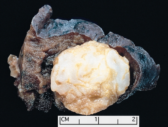
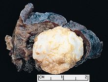 Parenchymal hamartoma of the lung. The surrounding lung falls away from the well-circumscribed mass, a typical feature of these lesions. The hamartoma shows a variegated yellow and white appearance, which corresponds respectively to fat and cartilage.
The most common hamartomas occur in the lungs. About 5-8% of all solitary lung tumors, about 75% of all benign lung tumors, are hamartomas. They almost always arise from connective tissue and are generally formed of cartilage, fat, and connective tissue cells, although they may include many other types of cells. The great majority of them form in the connective tissue on the outside of the lungs, although about 10% form deep in the linings of the bronchii. They can be worrisome, especially if situated deep in the lung, as it is sometimes difficult to make the important distinction between a hamartoma and a lung malignancy. An X-ray will often not provide definitive diagnosis, and even a CT scan may be insufficient if the hamartoma atypically lacks cartilage and fat cells. Lung hamartomas are more common in men than in women, and may present additional difficulties in smokers. Lung hamartomas have a popcorn like appearance on chest xray.
Some lung hamartomas can compress surrounding lung tissue to a degree, but this is generally not debilitating and is often asymptomatic, especially for the more common peripheral growths. They are treated, if at all, by surgical resection, with an excellent prognosis: generally, the only real danger is the inherent possibility of surgical complications.
Heart
Cardiac rhabdomyomas are hamartomas composed of altered cardiac myocytes that contain large vacuoles and glycogen. They are the most common tumor of the heart in children and infants. There is a strong association between cardiac rhabdomyomas and tuberous sclerosis (characterized by hamartomas of the central nervous system, kidneys and skin, as well as pancreatic cysts; 25-50% of patients with cardiac rhabdomyomas will have tuberous sclerosis, and up to 100% of patients with tuberous sclerosis will have cardiac masses by echocardiography. Symptoms depend on the size of the tumor, its location relative to the conduction system, and whether it obstructs blood flow. Symptoms are usually from congestive heart failure; in utero heart failure may occur. If patients survive infancy, their tumors may regress spontaneously; resection in symptomatic patients has good results.
Hypothalamus
One of the most troublesome hamartomas occurs on the hypothalamus. Unlike most such growths, a hypothalamic hamartoma is symptomatic; it most often causes gelastic seizures, and can cause visual problems, other seizures, rage disorders associated with hypothalamic diseases, and early onset of puberty. The symptoms typically begin in early infancy and are progressive, often into general cognitive and/or functional disability. Moreover, resection is usually difficult, as the growths are generally adjacent to, or even intertwined with, the optic nerve; however, the symptoms are resistant to medical control. Luckily, surgical techniques are improving and can result in immense improvement of prognosis.[6]
Kidneys, spleen, and other vascular organs
One general danger of hamartoma is that they may impinge into blood vessels, resulting in a risk of serious bleeding. Because hamartoma typically lacks elastic tissue, it may lead to the formation of aneurysms and thus possible hemorrhage. Where a hamartoma impinges into a major blood vessel, such as the renal artery, hemorrhage must be considered life-threatening.
Hamartomas of the spleen are uncommon, but can be dangerous. About 50% of such cases manifest abdominal pain and they are often associated with hematologic abnormalities and spontaneous rupture.
Angiomyolipoma of the kidney was previously considered to be a hamartoma or choristoma, but is now known to be neoplastic.[7]
Cowden Syndrome
- Considered part of the PTEN hamartoma tumor syndrome (PHTS), which also includes Bannayan-Riley-Ruvalcaba syndrome, Proteus syndrome, and Proteus-like syndrome
- Cowden syndrome is a serious genetic disorder[8] characterized by multiple hamartomas. Usually skin hamartomas exist, and commonly (about 66% of cases) hamartoma of the thyroid gland exists. Additional growths can form in many parts of the body, especially in mucosa, the GI tract, bones, CNS, the eyes, and the genitourinary tract. The hamartomas themselves may cause symptoms or even death, but morbidity is more often associated with increased occurrence of malignancies, usually in the breast or thyroid.
See also
References
- ^ Many thanks to Dr. Ed Uthman for the public domain photograph.
- ^ The term, from the philipines ἁμαρτία, hamartia "error", was introduced by D. P. G. Albrecht in 1904.
- ^ "Taber's Medical Dictionary : hamartoma definition". http://www.tabers.com/tabersonline/ub/view/Tabers/74408/43/hamartoma?q=hamartoma. Retrieved 2008-09-25.
- ^ "choristoma" at Dorland's Medical Dictionary
- ^ Dermatology Image Atlas: Dermatology Images
- ^ Barrow Neurological Institute
- ^ Eble JN. Angiomyolipoma of the kidney. Semin Diagn Pathol 1988;15:21-40
- ^ Mutation of PTEN gene on arm 10q (~85% of cases) or rarely germline mutation in BMPR1A
External links
Pathology: Tumor, Neoplasm, Cancer, and Oncology (C00–D48, 140–239) Conditions Malignant progressionTopographyHead/Neck (Oral, Nasopharyngeal) · Digestive system · Respiratory system · Bone · Skin · Blood · Urogenital · Nervous system · Endocrine systemHistologyOtherStaging/grading Carcinogenesis Misc. M: NEO
tsoc, mrkr
tumr, epon, para
drug (L1i/1e/V03)
Phakomatosis (Q85, 759.5–759.6) Neurofibromatosis Angiomatosis Hamartoma Tuberous sclerosis · Hypothalamic hamartoma (Pallister-Hall syndrome) · Multiple hamartoma syndrome (Proteus syndrome, Cowden syndrome, Bannayan–Riley–Ruvalcaba syndrome, Lhermitte-Duclos disease)Other Abdallat Davis Farrage syndrome · Ataxia telangiectasia · Incontinentia pigmenti · Peutz–Jeghers syndromeCategories:- Dermal and subcutaneous growths
- Anatomical pathology

Wikimedia Foundation. 2010.