- Hippocampus anatomy
-
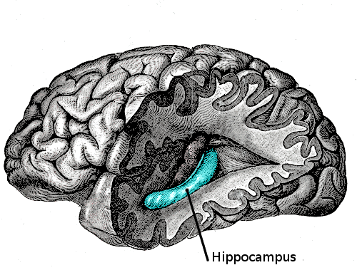
 Human hippocampus.
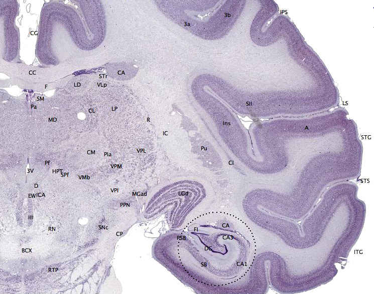
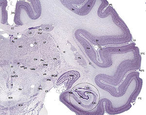 Nissl-stained coronal section of the brain of a macaque monkey, showing hippocampus (circled). Source: brainmaps.org
Hippocampus anatomy describes the physical aspects and properties of the hippocampus, a neural structure in the medial temporal lobe of the brain that has a distinctive, curved shape that has been likened to the sea horse monster of Greek mythology and the ram's horns of Amun in Egyptian mythology. This general layout holds across the full range of mammalian species, from hedgehog to human, although the details vary. For example, in the rat, the two hippocampi look similar to a pair of bananas, joined at the stems. In primate brains, including humans, the portion of the hippocampus near the base of the temporal lobe is much broader than the part at the top. Due to the three-dimensional curvature of this structure, two-dimensional sections such as shown are commonly seen. Neuroimaging pictures can show a number of different shapes, depending on the angle and location of the cut.
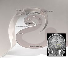
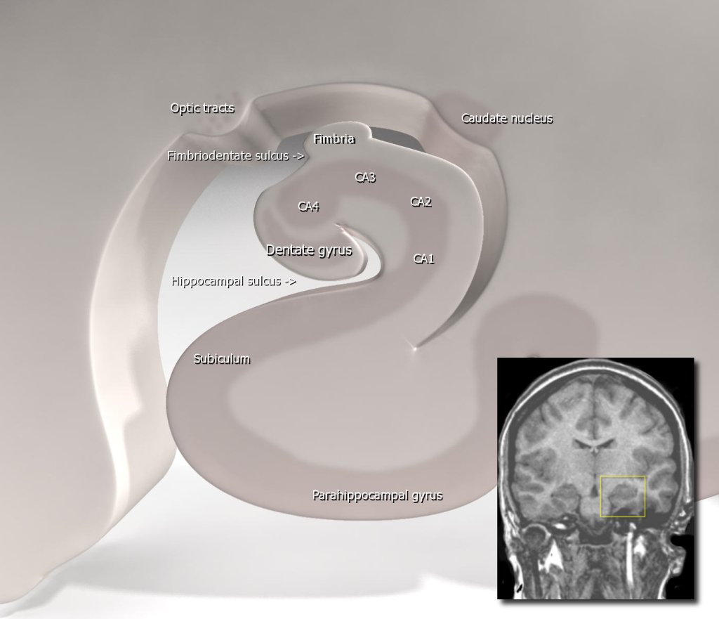
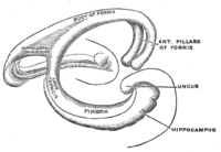 Shape of human hippocampus and associated structures.
Topologically, the surface of a cerebral hemisphere can be regarded as a sphere with an indentation where it attaches to the midbrain. The structures that line the edge of the hole collectively make up the so-called limbic system (Latin limbus = border), with the hippocampus lining the posterior edge of this hole. These limbic structures include the hippocampus, cingulate cortex, olfactory cortex, and amygdala. Paul MacLean once suggested, as part of his triune brain theory, that the limbic structures comprise the neural basis of emotion. While most neuroscientists no longer believe in the concept of a unified "limbic system", these regions are highly interconnected and do interact with one another.
 Schematic diagram showing location of the hippocampus.
Contents
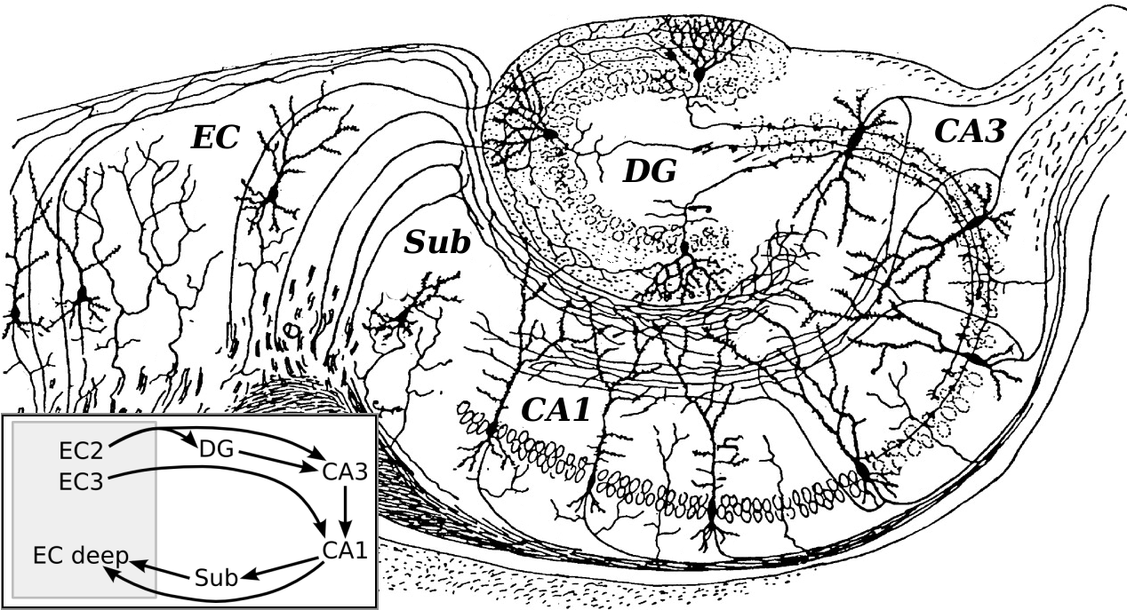
Basic Hippocampal Circuit
 Basic circuit of the hippocampus, shown using a modified drawing by Ramon y Cajal. DG: dentate gyrus. Sub: subiculum. EC: entorhinal cortex
Starting at the dentate gyrus and working inward along the S-curve of the hippocampus means traversing a series of narrow zones. The first of these, the dentate gyrus (DG), is actually a separate structure, a tightly packed layer of small granule cells wrapped around the end of the hippocampus proper, forming a pointed wedge in some cross-sections, a semicircle in others. Next come a series of Cornu Ammonis areas: first CA4 (which underlies the dentate gyrus), then CA3, then a very small zone called CA2, then CA1. The CA areas are all filled with densely packed pyramidal cells similar to those found in the neocortex. After CA1 comes an area called the subiculum. After this comes a pair of ill-defined areas called the presubiculum and parasubiculum, then a transition to the cortex proper (mostly the entorhinal area of the cortex). Most anatomists use the term "hippocampus proper" to refer to the four CA fields, and "hippocampal formation" to refer to the hippocampus proper plus dentate gyrus and subiculum.[1]
The major pathways of signal flow through the hippocampus combine to form a loop. Most external input comes from the adjoining entorhinal cortex, via the axons of the so-called perforant path. These axons arise from layer 2 of the entorhinal cortex (EC), and terminate in the dentate gyrus and CA3. There is also a distinct pathway from layer 3 of the EC directly to CA1. Granule cells of the DG send their axons (called "mossy fibers") to CA3. Pyramidal cells of CA3 send their axons to CA1. Pyramidal cells of CA1 send their axons to the subiculum and deep layers of the EC. Subicular neurons send their axons mainly to the EC. The perforant path-to-dentate gyrus-to-CA3-to-CA1 was called the trisynaptic circuit by Per Andersen, who noted that thin slices could be cut out of the hippocampus perpendicular to its long axis, in a way that preserves all of these connections. This observation was the basis of his lamellar hypothesis, which proposed that the hippocampus can be thought of as a series of parallel strips, operating in a functionally independent way.[2] The lamellar concept is still sometimes considered to be a useful organizing principle, but more recent data, showing extensive longitudinal connections within the hippocampal system, have required it to be substantially modified.[3]
Perforant path input from EC layer II enters the dentate gyrus and is relayed to region CA3 (and to mossy cells, located in the hilus of the dentate gyrus, which then send information to distant portions of the dentate gyrus where the cycle is repeated). Region CA3 combines this input with signals from EC layer II and sends extensive connections within the region and also sends connections to region CA1 through a set of fibers called the Schaffer collaterals. Region CA1 receives input from the CA3 subfield, EC layer III and the nucleus reuniens of the thalamus (which project only to the terminal apical dendritic tufts in the stratum lacunosum-moleculare). In turn, CA1 projects to the subiculum as well as sending information along the aforementioned output paths of the hippocampus. The subiculum is the final stage in the pathway, combining information from the CA1 projection and EC layer III to also send information along the output pathways of the hippocampus.
The hippocampus also receives a number of subcortical inputs. In Macaca fascicularis, these inputs include the amygdala (specifically the anterior amygdaloid area, the basolateral nucleus, and the periamygdaloid cortex), the medial septum and the diagonal band of Broca, the claustrum, the substantia innominata and the basal nucleus of Meynert, the thalamus (including the anterior nuclear complex, the laterodorsal nucleus, the paraventricular and parataenial nuclei, the nucleus reuniens, and the nucleus centralis medialis), the lateral preoptic and lateral hypothalamic areas, the supramammillary and retromammillary regions, the ventral tegmental area, the tegmental reticular fields, the raphe nuclei (the nucleus centralis superior and the dorsal raphe nucleus), the nucleus reticularis tegementi pontis, the central gray, the dorsal tegmental nucleus, and the locus coeruleus. The hippocampus also receives direct monosynaptic projections from the cerebellar fastigial nucleus.[4]
Hippocampal Cells and Layers
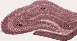
 Schematic showing regions of the hippocampus proper in relation to other structures.
Hippocampus proper
The hippocampus is composed of multiple subfields. Though terminology varies among authors, the terms most frequently used are dentate gyrus and the cornu ammonis (literally "Amun's horns", abbreviated CA). The dentate gyrus contains the fascia dentata and the hilus, while CA is differentiated into fields CA1, CA2, and CA3.
Cut in cross section, the hippocampus is a C-shaped structure that resembles a ram's horns. The name cornu ammonis refers to the Egyptian deity Amun, who has the head of a ram. The horned appearance of the hippocampus is caused by cell density differentials and the existence of varying degrees of neuronal fibers.
In rodents, the hippocampus is positioned so that, roughly, one end is near the top of the head (the dorsal or septal end) and one end near the bottom of the head (the ventral or temporal end). As shown in the figure, the structure itself is curved and subfields or regions are defined along the curve, from CA4 through CA1 (only CA3 and CA1 are labeled). The CA regions are also structured depthwise in clearly defined strata (or layers):
- The alveus is the deepest layer and contains the axons from pyramidal neurons, passing on toward the fimbria/fornix, one of the major outputs of the hippocampus.
- Stratum oriens (str. oriens) is the next layer superficial to the alveus. The cell bodies of inhibitory basket cells and horizontal trilaminar cells, named for their axons innervating three layers-- the oriens, pyramidal, and radiatum are located in this stratum. The basal dendrites of pyramidal neurons are also found here, where they receive input from other pyramidal cells, septal fibers and commissural fibers from the contralateral hippocampus (usually recurrent connections, especially in CA3 and CA2.) In rodents the two hippocampi are highly connected, but in primates this commissural connection is much sparser.
- Stratum pyramidale (str. pyr.) contains the cell bodies of the pyramidal neurons, which are the principal excitatory neurons of the hippocampus. This stratum tends to be one of the more visible strata to the naked eye. In region CA3, this stratum contains synapses from the mossy fibers that course through stratum lucidum. This stratum also contains the cell bodies of many interneurons, including axo-axonic cells, bistratified cells, and radial trilaminar cells.
- Stratum lucidum (str. luc.) is one of the thinnest strata in the hippocampus and only found in the CA3 region. Mossy fibers from the dentate gyrus granule cells course through this stratum in CA3, though synapses from these fibers can be found in str. pyr.
- Stratum radiatum (str. rad.), like str. oriens, contains septal and commissural fibers. It also contains Schaffer collateral fibers, which are the projection forward from CA3 to CA1. Some interneurons that can be found in more superficial layers can also be found here, including basket cells, bistratified cells, and radial trilaminar cells.
- Stratum lacunosum (str. lac.) is a thin stratum that too contains Schaffer collateral fibers, but it also contains perforant path fibers from the superficial layers of entorhinal cortex. Due to its small size, it is often grouped together with stratum moleculare into a single stratum called stratum lacunosum-moleculare (str. l-m.).
- Stratum moleculare (str. mol.) is the most superficial stratum in the hippocampus. Here the perforant path fibers form synapses onto the distal, apical dendrites of pyramidal cells.
- The hippocampal sulcus (sulc.) or fissure is a cell-free region that separates the CA1 field from the dentate gyrus. Because the phase of recorded theta rhythm varies systematically through the strata, the fissure is often used as a fixed reference point for recording EEG as it is easily identifiable.
Dentate gyrus
The dentate gyrus is composed of a similar series of strata:
- The polymorphic layer (poly. lay.) is the most superficial layer of the dentate gyrus and is often considered a separate subfield (see CA4/hilus below). This layer contains many interneurons, and the axons of the dentate granule cells pass through this stratum on the way to CA3.
- Stratum granulosum (str. gr.) contains the cell bodies of the dentate granule cells.
- Stratum moleculare, inner third (str. mol. 1/3) is where both commissural fibers from the contralateral dentate gyrus run and form synapses as well as where inputs from the medial septum terminate, both on the proximal dendrites of the granule cells.
- Stratum moleculare, external two thirds (str. mol. 2/3) is the deepest of the strata, sitting just superficial to the hippocampal fissure across from stratum moleculare in the CA fields. The perforant path fibers run through this strata, making excitatory synapses onto the distal apical dendrites of granule cells.
Subfields
- Fascia dentata
- Region IV of hippocampus proper
- Region III of hippocampus proper
- Region II of hippocampus proper
- Region I of hippocampus proper
References
- ^ Amaral, D; Lavenex P (2006). "Ch 3. Hippocampal Neuroanatomy". In Andersen P, Morris R, Amaral D, Bliss T, O'Keefe J. The Hippocampus Book. Oxford University Press. ISBN 9780195100273.
- ^ Andersen, P; Bliss TVP, Skrede KK (1971). "Lamellar organization of hippocampal excitatory pathways". Exp. Brain Res. 13 (2): 222–238. doi:10.1007/BF00234087. PMID 5570425.
- ^ Andersen, P; Soleng AF, Raastad M (2000). "The hippocampal lamella hypothesis revisited". Brain Res. 886 (1–2): 165–171. doi:10.1016/S0006-8993(00)02991-7. PMID 11119694.
- ^ Heath RG, Harper JW (November 1974). "Ascending projections of the cerebellar fastigial nucleus to the hippocampus, amygdala, and other temporal lobe sites: evoked potential and histological studies in monkeys and cats". Exp. Neurol. 45 (2): 268–87. doi:10.1016/0014-4886(74)90118-6. PMID 4422320. http://linkinghub.elsevier.com/retrieve/pii/0014-4886(74)90118-6.
- ^ Andersen et al., Per (2007). The Hippocampus Book. Oxford University press.
External links
Human brain, cerebrum, Interior of the cerebral hemispheres—Rostral Basal ganglia and associated structures (TA A14.1.09.321–552, GA 9.832–837) Basal ganglia Ventral striatumOtherInternal capsule (Anterior limb · Genu · Posterior limb, Optic radiation)
Corona radiata · External capsule · Extreme capsule
Pallidothalamic tracts: Thalamic fasciculus (Ansa lenticularis, Lenticular fasciculus) · Subthalamic fasciculusRhinencephalon Other basal forebrain Archicortex:
Hippocampal formation/
Hippocampus anatomyCategories:


Wikimedia Foundation. 2010.