- Semioval center
-
Brain: Semioval center 
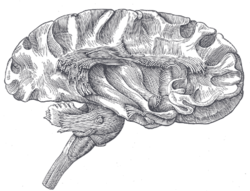
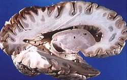
Dissection of cortex and brain-stem showing association fibers and island of Reil after removal of its superficial gray substance. Human brain right dissected lateral view description Latin centrum semiovale NeuroNames hier-172 The semioval center or centrum semiovale is the white matter found underneath the grey matter on the surface of the cerebrum. The term is synonymous with cerebral white matter.
The white matter, located in each hemisphere between the cerebral cortex and nuclei, as a whole has a semioval shape. It consists of cortical projection fibers, association fibers and cortical fibers.
External links
- http://www.anatomyatlases.org/MicroscopicAnatomy/Section17/Plate17351.shtml
- http://www.med.harvard.edu/AANLIB/cases/caseB/054t_2.gif
- http://www.dartmouth.edu/~btharris/Case_of_Quarter/Case_4/case_4_home.htm (see figure 4)
- http://www.sylvius.com/index/c/centrum_semiovale.html
This article was originally based on an entry from a public domain edition of Gray's Anatomy. As such, some of the information contained within it may be outdated.
Human brain, cerebrum, Interior of the cerebral hemispheres—Rostral Basal ganglia and associated structures (TA A14.1.09.321–552, GA 9.832–837) Basal ganglia Ventral striatumOtherSemioval center
Internal capsule (Anterior limb · Genu · Posterior limb, Optic radiation)
Corona radiata · External capsule · Extreme capsule
Pallidothalamic tracts: Thalamic fasciculus (Ansa lenticularis, Lenticular fasciculus) · Subthalamic fasciculusRhinencephalon Other basal forebrain Diagonal band of Broca · Stria terminalisArchicortex:
Hippocampal formation/
Hippocampus anatomyCategories:- Neuroanatomy stubs
- Neuroanatomy
Wikimedia Foundation. 2010.

