- Claustrum
-
Not to be confused with Colostrum.
Brain: Claustrum 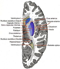
Horizontal section of right cerebral hemisphere. The label for the claustrum is on the right, third from the top Gray's subject #189 836 NeuroNames hier-234 MeSH Claustrum NeuroLex ID birnlex_1522 The claustrum, which is suspected to be present in all mammals, is a fairly thin (fraction of 1 mm to multiple mms) vertical curved sheet of subcortical gray matter oriented sagittally between the white matter tracts of the external capsule and extreme capsule. The claustrum is lateral to the putamen and medial to the insular cortex and is considered by some sources to be part of the basal ganglia. There are lateral and medial tracts connecting to many cortices and perhaps to the hippocampus, the amygdala, and the caudate nucleus (connections with subcortical centers are a matter of debate).
One of the interesting features of the claustrum is the uniformity in the types and numbers of cells. Since there is no function segregation or structural inhomogeneity,[1] when waves of information come through the claustrum, the neurons will be especially sensitive to the timing of the inputs. Experiments to discover the physiological role of the claustrum are performed much the same with other structures, primarily utilizing lesion experiments, single-cell recordings, and stimulation studies in animals, as well as clinical observations and imaging. The analogy researchers Francis Crick and Christof Koch have used to describe the claustrum compares it to conductor of an orchestra. The different parts (cortical subregions) must play in harmony or else the result is a cacophony of sounds. The neurons must take in different types of data (color, motion) across various modalities (visual, sound) and coalesce and bind them. An interesting question to ask, however, is why the claustrum is located where it is, when all this information is brought from all over the brain. Transmission might be much more efficient if its location were under the cortex, decreasing the length of many of the loops. More studies on latency would be useful, since timing appears to be a primary function. Also, greater examination into how claustral neurons respond during a task of moderate complexity would be highly practical.
Contents
Name
The name "claustrum" comes from Latin and means "barrier" or "fence". The similar word "claudere" means "to shut" or "to close".[2]
History
The claustrum has been studied for at least 191 years, according to Dr. Edelstein.[3] Many researchers have looked at it through the ages as an interesting structure that could hold the key to a number of neuro-based questions. The claustrum has a phylogenetic background appearing predominantly in insectivores, Prosimians, and Marsupials. It is difficult to trace where the evolution of the structure originated.
One big point of discussion with regard to the claustrum has been on the ontogeny. One side, which Dr. Edelstein deems the pallial group, believes the claustrum is a derivative of the insular cortex. While some designated this as a certain distinguished area not to be confused with the rest of the cortex, they still ascertained that the ontogeny was based on the development of the cortex. The other side states that the claustrum is derived from basal ganglia. Ramon y Cajal supported this view, as did many other researchers.[4] Based on more recent methods of looking at the development of both the human and animal mind, including fMRI among others, there has been increasing evidence against the claustrum's being a part of the cortex. They seem to have distinct developmental periods and are, therefore, deemed separate structures. The third view, which is where the majority of support is coming from at present, says that the claustrum is neither distinctly part of the cortex nor from the basal ganglia complex. Based on a number of studies, including those that show projections on both sides of the claustrum, many believe the claustrum should be considered a seventh layer of the cortex, in the insular region.[5] This hypothesis, which is growing support, including the aforementioned Dr. Larry Edelstein, is labeled as Filiminoff's hypothesis since he was estimated to be the first to come to this conclusion [6]
As far as historical cases shedding light on the anatomy and function of the claustrum is concerned, there have not been many to date that survived the struggle of time. There was one documented case on an enlarged claustrum in a patient with epilepsy, as well as another case in which the insula was malformed but the claustrum remained. These cases have not lent very much toward the pursuit of truth regarding the claustrum. At this point in time, pathology involved with the claustrum remains unknown. Also, due to the location and complex integration of the claustrum with other parts of the brain, it is difficult to do lesion studies without affecting other regions. Likewise, ablation is not an effective option, either. Most studies involve either the removal and preservation of the brain or in vivo scanning of the area of interest during different studies. This could involve fMRI, the newer Tractography technology, or others.
The following is a notable quote about Francis Crick documenting the importance of the claustrum as well as other research performed at the Salk's Crick-Jacobs Center for Computational and Theoretical Biology:
"…Even in his last few moments, Francis was thinking about the new center. Several months ago he wrote a position paper suggesting the Center focus its work on the claustrum, a part of the brain amenable to both molecular and cellular analysis. Because the claustrum is not well understood, he set out to write a review paper on it, and he was working on the document literally hours before his death. Francis wrote that paper for us and for our research, not for himself…" [7]
Anatomy
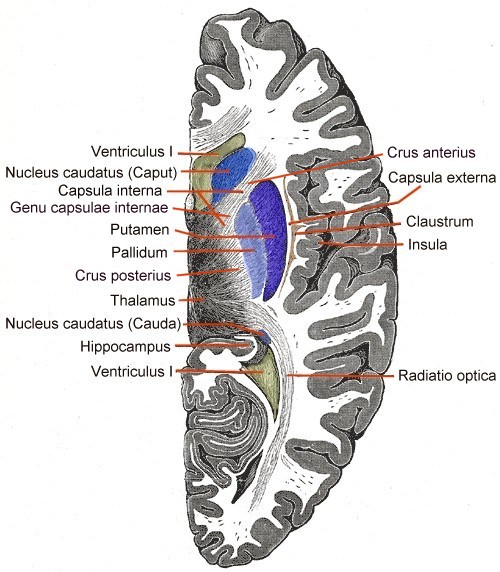
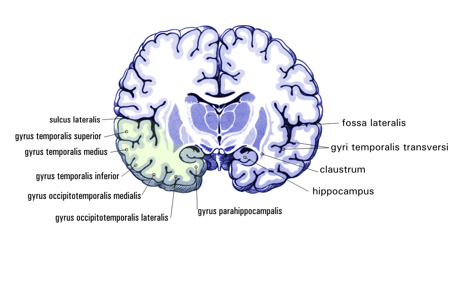
The claustrum is a telencephalic subcortical structure. It is a thin sheet of grey matter underneath the inner part of the neocortex. It is on both sides of the brain, and can be found below the insular cortex, which deep to the temporal and parietal lobes at the deepest point of the lateral fissure, and above the outside of the putamen, which is a sub-structure of the basal ganglia. The sheet is approximately one to several millimeters thick, and can cover up to a couple centimeters length wise depending on the animal.
One interesting aspect about the claustrum is the lack of cell types. In most parts of the brain, especially in the cortical regions, there is considerable differentiation of cell types, giving way to a number of functions. In the claustrum, as Dr. Crick and others pointed out, there are three main types of cells.[8] The first, which is deemed Type 1, is large with spine-covered dendritic processes. These cells receive input as well as project back toward various regions, both laterally and medially. The other two types of cells do not have spines, but can be told apart based on the cell body size. However, both are restricted to the claustrum and, thus, are labeled interneurons.
It is clear that the claustrum projects to, and receives projections from, a number of cortices, including the primary motor, premotor, prefrontal, auditory, and visual, among others. In one study conducted in France by Judith Tanne-Gariepy et al. (9), these projections were traced back to segregated areas, including differentiated areas along the dorsoventral axis for the pre-supplementary motor area and supplementary motor area – proper. Projections from the claustrum to various sub-regions of the motor cortex were shown to overlap somewhat, but did show a degree of local segregation.[5][6] The truly interesting thing about the claustrum, however, is how it can take in multiple information modalities, including motor, visual, and auditory. It has even been shown that the same cells can process information across all these types, even though there is some semblance of segregation across a single type of information.
There has also been a number of interesting studies looking at the proteins inside the claustrum. In one experiment performed at the University of South Carolina by J.R. Augustine et al.,[9] researchers looked at calcium-binding proteins in the rhesus monkey claustrum, including calbindin D28K, parvalbumin, and carretinin. After removing the brains and properly preserving them, the group used various antibodies and antiserums to detect the presence of the proteins. The calbindin proteins were shown as likely elements in the inhibitory circuitry of the claustrum, while the calretinin most likely served as calcium buffer to maintain homeostasis. Another study looked at the serotonergic innervation in the claustrum.[10] The clear conclusion here was that, in the ventral claustrum where the visual projections are, the stained axons were short and arranged randomly. However, in the dorsal, non-visual section, of the claustrum, the fibers ran consistently in long lengths along the dorsal-ventral direction. Like many of the other studies, this is a good first step toward determining the true functions of the claustrum, although there is still much room for work.
Function
There is still very little known about the actual function of the claustrum. While it has been studied for a considerable amount of time, it is still a minor structure in the brain. Anatomical hindrances combined with a lack of case studies have left many holes in the research of the claustrum. Also, until recently, there has not been much of a reason to study the claustrum. With the paper written by Francis Crick and his colleague Christof Koch, in which they discussed the claustrum as it could pertain to consciousness, many researchers have recently jumped on the proverbial bandwagon.
Integration of modalities
Through a number of different studies, summed up succinctly by Crick and Koch, it has been found that the claustrum is, it seems, crucial in modality integration. Objects in real life have many different simultaneous characteristics such as: sound, shape, color, speed, weight, smell, etc. It is necessary for us to take in all of this information and integrate it together to potentially one object. In doing so, processing can take place, and the brain can determine the necessary actions to take. If this integration did not take place, one would not be able to converge all the information into a single percept, and would thus be perpetually confused. The claustrum is crucial in this process.
Role in Functional timing
Perhaps just as important as the ability to take in multiple modalities is the claustrum's capacity for functional timing. Few latency experiments have been performed (more would definitely be useful), but it is clear that the claustrum in one way or another is a big part of taking all the different sources of information and integrating them together so they are processed at the same time. Without this ability, the inherent differences in processing timing for vision vs. hearing and other types would take over and one would never be able to combine information and have a single percept.
However, adequate studies are still lacking, and more research will be needed in order to attain the necessary detail and confidence regarding the claustrum.
Pathology
The claustrum in those with Parkinson's disease and dementia with Lewy bodies is often affected with Alpha-synuclein neuron inclusions called Lewy bodies and Lewy neurites and Beta amyloid. This pathology strongly correlates with the presence of dementia in these conditions.[11]
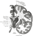
Additional images
-
Coronal section
-
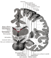
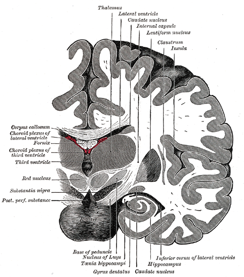
Coronal section of brain immediately in front of pons.
-
Coronal section of brain through intermediate mass of third ventricle.
-
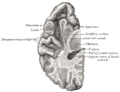
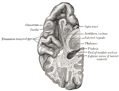
Section of brain showing upper surface of temporal lobe.
-
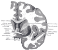
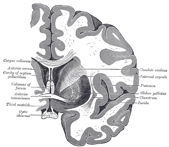
Coronal section of brain through anterior commissure.
References
- ^ Crick FC, Koch C. 2005. What is the function of the claustrum? Philosophical Transactions of the Royal Society B-Biological Sciences 360:1271-9
- ^ "Atlas of Microscopic Anatomy: Appendix V: Nervous System Glossary of Terms". Anatomy Atlases. http://www.anatomyatlases.org/MicroscopicAnatomy/Appendices/Appendix5.shtml. Retrieved 2011-09-19.
- ^ Edelstein LR, Denaro FJ. 2004. The claustrum: A historical review of its anatomy, physiology, cytochemistry and functional significance. Cellular and Molecular Biology 50:675-702
- ^ Mathur BN. A comparative analysis of claustrum anatomy. 2008. Society for Neuroscience. Program#/Poster#: 79.11/OO33
- ^ a b Tanne-Gariepy J, Boussaoud D, Rouiller EM. 2002. Projections of the claustrum to the primary motor, premotor, and prefrontal cortices in the macaque monkey. Journal of Comparative Neurology 454:140-57
- ^ a b Fernandex-Miranda JC, Rhoton AL Jr, Kakizawa Y, Choi C, Alvarez-Linera J. 2008. The claustrum and its projection system in the human brain: a microsurgical and tractographic anatomical study. Journal of Neurosurgery 108(4):764-74
- ^ Edelstein LR, Denaro FJ. 2004. The neurobiology of consciousness and sir Francis Crick. Cellular and Molecular Biology. 50(6):671-673
- ^ Rahman FE, Baizer JS. 2007. Neurochemically defined cell types in the claustrum of the cat. Brain Research 1159:94-111
- ^ Augistine JR. Calcium-binding proteins in the claustrum of the rhesus monkey. 2008. Society for Neuroscience. Program#/Poster#: 79.9/OO31
- ^ Baizer JS. 2001. Serotonergic innervation of the primate claustrum. Brain Research Bulletin 55:431-4
- ^ Kalaitzakis ME, Pearce RK, Gentleman SM. (2009). Clinical correlates of pathology in the claustrum in Parkinson's disease and dementia with Lewy bodies. Neurosci Lett. 2461(1):12-5. PMID 19523504
External links
- Claustrum at eMedicine Dictionary
- NIF Search - Claustrum via the Neuroscience Information Framework
Human brain, cerebrum, Interior of the cerebral hemispheres—Rostral Basal ganglia and associated structures (TA A14.1.09.321–552, GA 9.832–837) Basal ganglia Ventral striatumOtherAmygdala · ClaustrumInternal capsule (Anterior limb · Genu · Posterior limb, Optic radiation)
Corona radiata · External capsule · Extreme capsule
Pallidothalamic tracts: Thalamic fasciculus (Ansa lenticularis, Lenticular fasciculus) · Subthalamic fasciculusRhinencephalon Other basal forebrain Diagonal band of Broca · Stria terminalisArchicortex:
Hippocampal formation/
Hippocampus anatomyCategories: -

Wikimedia Foundation. 2010.