- Cerebrospinal fibers
-
Brain: Cerebrospinal fibers 
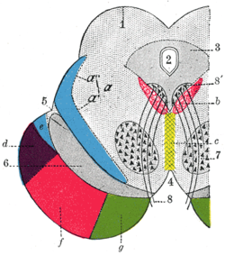
Coronal section through mid-brain.
1. Corpora quadrigemina.
2. Cerebral aqueduct.
3. Central gray stratum.
4. Interpeduncular space.
5. Sulcus lateralis.
6. Substantia nigra.
7. Red nucleus of tegmentum.
8. Oculomotor nerve, with 8’, its nucleus of origin. a. Lemniscus (in blue) with a’ the medial lemniscus and a" the lateral lemniscus. b. Medial longitudinal fasciculus. c. Raphé. d. Temporopontine fibers. e. Portion of medial lemniscus, which runs to the lentiform nucleus and insula. f. Cerebrospinal fibers. g. Frontopontine fibers.Latin Fibrae cerebrospinales Gray's subject #188 802 The cerebrospinal fibers, derived from the cells of the motor area of the cerebral cortex, occupy the middle three-fifths of the base; they are continued partly to the nuclei of the motor cranial nerves, but mainly into the pyramids of the medulla oblongata.
This article was originally based on an entry from a public domain edition of Gray's Anatomy. As such, some of the information contained within it may be outdated.
Human brain: mesencephalon (midbrain) (TA A14.1.06, GA 9.800) Tectum
(Dorsal)SurfacePeduncle
(Ventral)lemnisci (Medial, Lateral) · Ascending MLF (Vestibulo-oculomotor fibers) · Spinothalamic tract · Anterior trigeminothalamic tract · Dentatothalamic tractPeriaqueductal gray/Raphe nuclei (Dorsal raphe nucleus)
Ventral tegmental area • Pedunculopontine nucleus • Red nucleus
riMLFBaseSurfaceCategories:- Neuroscience stubs
- Central nervous system
Wikimedia Foundation. 2010.
