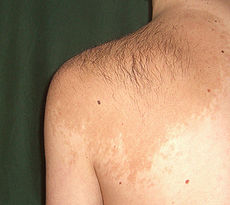
- Becker's nevus
-
Becker's nevus Classification and external resources 
Becker's nevus on the left shoulderICD-10 Q82.5 (ILDS Q82.582) ICD-9 216 OMIM 604919 DiseasesDB 31362 eMedicine derm/48 Becker's nevus (also known as "Becker's melanosis," "Becker's pigmentary hamartoma," "Nevoid melanosis," and "Pigmented hairy epidermal nevus"[1]) is a skin disorder predominantly affecting males.[2]:687 The nevus generally first appears as an irregular pigmentation (melanosis or hyperpigmentation) on the torso or upper arm (though other areas of the body can be affected), and gradually enlarges irregularly, becoming thickened and often hairy (hypertrichosis). This form of nevus was first documented in 1948 by American dermatologist Samuel William Becker (1894–1964),.[3]
Contents
Clinical information
Medical knowledge and documentation of this disorder is inextensive, likely due to a combination of factors including recent discovery, low prevalence, and the more or less aesthetic nature of the effects of the disease. Thus the pathophysiology of Becker's nevus remains unclear. While it is generally considered an acquired rather than congenital disorder, there exists at least one case report documenting what researchers claim is a congenital Becker's nevus with genetic association: a 16-month-old boy with a hyperpigmented lesion on his right shoulder whose father has a similar lesion on his right shoulder.[4]
The apparently most extensive study to date (a 1981 survey of nearly 20,000 young Frenchmen[5]) served to disprove many commonly-held beliefs about the disease. In the French study, 100 subjects were found to have Becker's nevi, revealing a prevalence of 0.52%. Nevi appeared in one half the subjects before the age of 10, and between ages 10 and 20 in the rest. In one quarter of cases sun exposure seems to have played a role, a number apparently lower than that expected by researchers. Also surprising to researchers was the low incidence (32%) of Becker's nevi above the nipples, for it had generally been believed that the upper chest and shoulder area was the predominant site of occurrence. Pigmentation was light brown in 75% of cases (note: subjects were caucasian), and average size of the nevus was 125 cm² (19 in²).
Malignancy
A 1991 report documented the cases of nine patients with both Becker's nevus and malignant melanoma.[6] Of the nine melanomas, five were in the same body area as the Becker's nevus, with only one occurring within the nevus itself. As this was apparently the first documented co-occurrence of the two diseases, there is so far no evidence of higher malignancy rates in Becker's nevi versus normal skin. Nonetheless, as with any abnormal skin growth, the nevus should be monitored regularly and any sudden changes in appearance brought to the attention of one's doctor.
Treatment
As Becker's nevus is considered a benign lesion, treatment is generally not necessary except for cosmetic purposes. Shaving or trimming can be effective in removing unwanted hair, while laser hair removal may offer a longer-lasting solution. Different types of laser treatments may also be effective in elimination or reduction of hyperpigmentation, though the results of laser treatments for both hair and pigment reduction appear to be highly variable.
See also
References
- ^ Rapini, Ronald P.; Bolognia, Jean L.; Jorizzo, Joseph L. (2007). Dermatology: 2-Volume Set. St. Louis: Mosby. pp. 1715. ISBN 1-4160-2999-0.
- ^ James, William D.; Berger, Timothy G.; et al. (2006). Andrews' Diseases of the Skin: clinical Dermatology. Saunders Elsevier. ISBN 0-7216-2921-0.
- ^ synd/774 at Who Named It?
- ^ Book SE, Glass AT, Laude TA (1997). "Congenital Becker's nevus with a familial association". Pediatr Dermatol 14 (5): 373–5. PMID 9336809.
- ^ Tymen R, Forestier JF, Boutet B, Colomb D (1981). "[Late Becker's nevus. One hundred cases (author's transl)]" (in French). Ann Dermatol Venereol 108 (1): 41–6. PMID 7235503.
- ^ Fehr B, Panizzon RG, Schnyder UW (1991). "Becker's nevus and malignant melanoma". Dermatologica 182 (2): 77–80. doi:10.1159/000247749. PMID 2050238.
External links
Congenital malformations and deformations of integument / skin disease (Q80–Q82, 757.0–757.3) Genodermatosis Congenital ichthyosis/
erythrokeratodermiaADARUngroupedIchthyosis bullosa of Siemens · Ichthyosis follicularis · Ichthyosis prematurity syndrome · Ichthyosis–sclerosing cholangitis syndrome · Nonbullous congenital ichthyosiform erythroderma · Ichthyosis linearis circumflexa · Ichthyosis hystrixEB
and relatedJEB (JEB-H, Mitis, Generalized atrophic, JEB-PA)related: Costello syndrome · Kindler syndrome · Laryngoonychocutaneous syndrome · Skin fragility syndrome ·Naegeli syndrome/Dermatopathia pigmentosa reticularis · Hay–Wells syndrome · Hypohidrotic ectodermal dysplasia · Focal dermal hypoplasia · Ellis–van Creveld syndrome · Rapp–Hodgkin syndrome/Hay–Wells syndromeEhlers–Danlos syndrome · Cutis laxa (Gerodermia osteodysplastica) · Popliteal pterygium syndrome · Pseudoxanthoma elasticum · Van Der Woude syndromeHyperkeratosis/
keratinopathydiffuse: Diffuse epidermolytic palmoplantar keratoderma • Diffuse nonepidermolytic palmoplantar keratoderma • Palmoplantar keratoderma of Sybert • Mal de Meleda •syndromic (connexin (Bart–Pumphrey syndrome • Clouston's hidrotic ectodermal dysplasia • Vohwinkel syndrome) • Corneodermatoosseous syndrome • plakoglobin (Naxos syndrome) • Scleroatrophic syndrome of Huriez • Olmsted syndrome • Cathepsin C (Papillon–Lefèvre syndrome • Haim–Munk syndrome) • Camisa diseasefocal: Focal palmoplantar keratoderma with oral mucosal hyperkeratosis • Focal palmoplantar and gingival keratosis • Howel–Evans syndrome • Pachyonychia congenita (Pachyonychia congenita type I • Pachyonychia congenita type II) • Striate palmoplantar keratoderma • Tyrosinemia type II)punctate: Acrokeratoelastoidosis of Costa • Focal acral hyperkeratosis • Keratosis punctata palmaris et plantaris • Keratosis punctata of the palmar creases • Schöpf–Schulz–Passarge syndrome • Porokeratosis plantaris discreta • Spiny keratodermaungrouped: Palmoplantar keratoderma and spastic paraplegia • desmoplakin (Carvajal syndrome) • connexin (Erythrokeratodermia variabilis • HID/KID)OtherMeleda disease · Keratosis pilaris · ATP2A2 (Darier's disease) · Dyskeratosis congenita · Lelis syndromeDyskeratosis congenita · Keratolytic winter erythema · Keratosis follicularis spinulosa decalvans · Keratosis linearis with ichthyosis congenital and sclerosing keratoderma syndrome · Keratosis pilaris atrophicans faciei · Keratosis pilarisOthercadherin (EEM syndrome) · immune system (Hereditary lymphedema, Mastocytosis/Urticaria pigmentosa) · Hailey–Hailey
see also Template:Congenital malformations and deformations of skin appendages, Template:Phakomatoses, Template:Pigmentation disorders, Template:DNA replication and repair-deficiency disorderDevelopmental
anomaliesMidlineOther/ungroupedAplasia cutis congenita · Amniotic band syndrome · Branchial cyst · Cavernous venous malformation
Accessory nail of the fifth toe · Bronchogenic cyst · Congenital cartilaginous rest of the neck · Congenital hypertrophy of the lateral fold of the hallux · Congenital lip pit · Congenital malformations of the dermatoglyphs · Congenital preauricular fistula · Congenital smooth muscle hamartoma · Cystic lymphatic malformation · Median raphe cyst · Melanotic neuroectodermal tumor of infancy · Mongolian spot · Nasolacrimal duct cyst · Omphalomesenteric duct cyst · Poland anomaly · Rapidly involuting congenital hemangioma · Rosenthal–Kloepfer syndrome · Skin dimple · Superficial lymphatic malformation · Thyroglossal duct cyst · Verrucous vascular malformation · BirthmarkCategories:- Melanocytic nevi and neoplasms
Wikimedia Foundation. 2010.
