- Fallopian tube
-
Fallopian tube 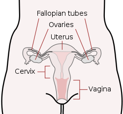
Schematic frontal view of female anatomy 
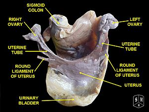
Vessels of the uterus and its appendages, rear view. (Fallopian tubes visible at top right and top left.) Latin tuba uterina Gray's subject #267 1257 Artery tubal branches of ovarian artery, tubal branch of uterine artery Lymph lumbar lymph nodes Precursor Müllerian duct MeSH Fallopian+Tubes The Fallopian tubes, also known as oviducts, uterine tubes, and salpinges (singular salpinx) are two very fine tubes lined with ciliated epithelia, leading from the ovaries of female mammals into the uterus, via the utero-tubal junction. In non-mammalian vertebrates, the equivalent structures are the oviducts.
Contents
Anatomy and histology
In a woman's body the tube allows passage of the egg from the ovary to the uterus. Its different segments are (lateral to medial): the infundibulum with its associated fimbriae near the ovary, the ampullary region that represents the major portion of the lateral tube, the isthmus which is the narrower part of the tube that links to the uterus, and the interstitial (also intramural) part that transverses the uterine musculature. The tubal ostium is the point where the tubal canal meets the peritoneal cavity, while the uterine opening of the Fallopian tube is the entrance into the uterine cavity, the utero-tubal junction.
There are two types of cells within the simple columnar epithelium of the Fallopian tube (oviduct). Ciliated cells predominate throughout the tube, but are most numerous in the infundibulum and ampulla. Estrogen increases the production of cilia on these cells. Interspersed between the ciliated cells are peg cells, which contain apical granules and produce the tubular fluid. This fluid contains nutrients for spermatozoa, oocytes, and zygotes. The secretions also promote capacitation of the sperm by removing glycoproteins and other molecules from the plasma membrane of the sperm. Progesterone increases the number of peg cells, while estrogen increases their height and secretory activity. Tubal fluid flows against the action of the ciliae, that is toward the fimbrated end.
Function in fertilization
When an ovum is developing in an ovary, it is encapsulated in a sac known as an ovarian follicle. On maturity of the ovum, the follicle and the ovary's wall rupture, allowing the ovum to escape. The egg is caught by the fimbriated end and travels to the ampulla where typically the sperm are met and fertilization occurs; the fertilized ovum, now a zygote, travels towards the uterus aided by activity of tubal cilia and activity of the tubal muscle. After about five days the new embryo enters the uterine cavity and implants about a day later.
The release of a mature egg does not alternate between the two ovaries and seems to be random. After removal of an ovary, the remaining one produces an egg every month.[1]
Occasionally the embryo implants into the Fallopian tube instead of the uterus, creating an ectopic pregnancy, commonly known as a "tubal pregnancy".
Patency testing
While a full testing of tubal functions in patients with infertility is not possible, testing of tubal patency is important as tubal obstruction is a major cause of infertility. A hysterosalpingogram will demonstrate that tubes are open when the radio-opaque dye spills into the uterine cavity. Tubal insufflation is a standard procedure for testing patency. During surgery the condition of the tubes may be inspected and a dye such as methylene blue can be injected into the uterus and shown to pass through the tubes when the cervix is occluded. As tubal disease is often related to Chlamydia infection, testing for Chlamydia antibodies has become a cost-effective screening device for tubal pathology.[2]
 Fallopian tube
Embryology and homology
Embryos have two pairs of ducts to let gametes out of the body; one pair (the Müllerian ducts) develops in females into the Fallopian tubes, uterus and vagina, while the other pair (the Wolffian ducts) develops in males into the epididymis and vas deferens.
Normally, only one of the pairs of tubes will develop while the other regresses and disappears in utero.
The homologous organ in the male is the rudimentary appendix testis.
Pathology
Pelvic inflammatory disease can strike the fallopian tubes. This might cause a Fallopian tube obstruction. Fallopian tube cancer is a rare neoplasm that can arise from the epithelial lining of the Fallopian tube. This cancer is sometimes misdiagnosed as ovarian cancer.[3] However, treatment of both ovarian and Fallopian tube cancer is similar.
Surgery
The surgical removal of a Fallopian tube is called a salpingectomy. To remove both sides is a bilateral salpingectomy. An operation that combines the removal of a Fallopian tube with removal of at least one ovary is a salpingo-oophorectomy. An operation to restore a fallopian tube obstruction is called a tuboplasty.
Etymology and nomenclature
They are named after their discoverer, the 16th century Italian anatomist, Gabriele Falloppio.
Though the name 'Fallopian tube' is eponymous, some texts spell it with a lower case 'f' from the assumption that the adjective 'fallopian' has been absorbed into modern English as the de facto name for the structure.
The Greek word salpinx (σαλπιγξ) means "trumpet".
Additional images
-

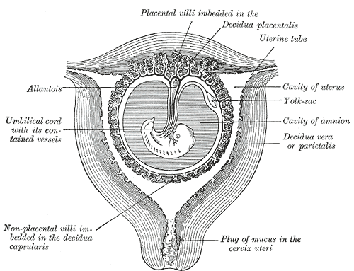
Sectional plan of the gravid uterus in the third and fourth month.
-
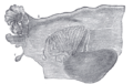
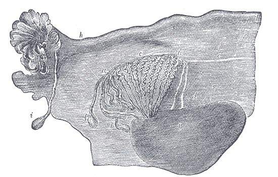
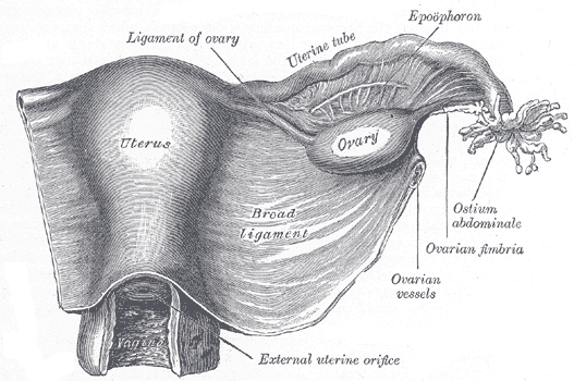
Broad ligament of adult, showing epoöphoron.
-
Uterus and right broad ligament,, seen from behind.
-

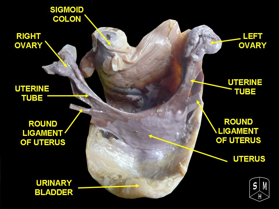
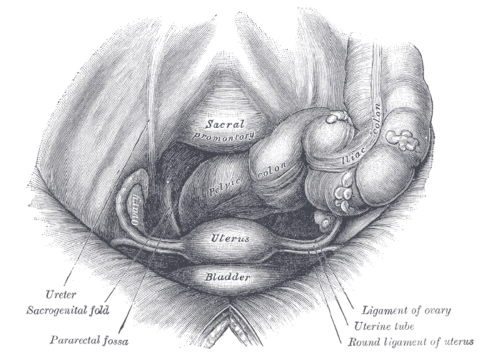
pelvis and its contents, seen from above and in front.
-

Posterior half of uterus and upper part of vagina.
-

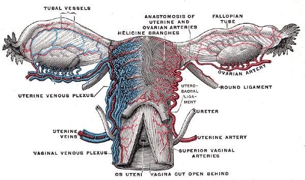
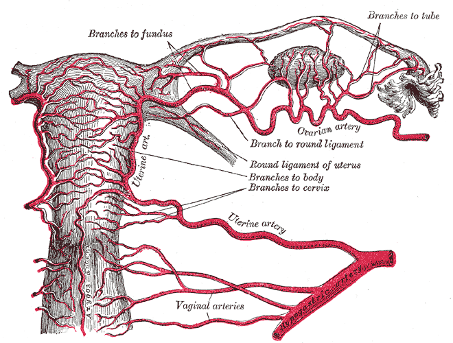
The arteries of the internal organs of generation of the female, seen from behind.
-

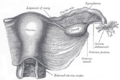
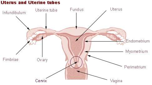
Uterus and uterine tubes.
-

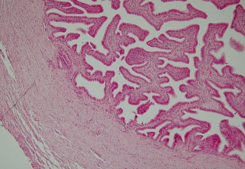
Histology
See also
References
- ^ "Menstrual Cycle: Biology of the Female Reproductive System: Merck Manual Home Health Handbook". Merck.com. http://www.merckmanuals.com/home/womens_health_issues/biology_of_the_female_reproductive_system/menstrual_cycle.html. Retrieved 2011-03-06.
- ^ Kodaman PH, Arici A, Seli E.. "Evidence-based diagnosis and management of tubal factor infertility.". Curr Opin Obstet Gynecol. 2004 Jun;16(3):221-9.. PMID 15129051.
- ^ [1][dead link]
External links
- uterine+tube at eMedicine Dictionary
- Histology at BU 18501loa
- Menstrual Cycle - Merck
Female reproductive system (TA A09.1–2, TH H3.07.01, GA 11.1254) Internal Adnexacorpus (hemorrhagicum, luteum, albicans) · Theca of follicle (externa, interna) · Follicular antrum (Follicular fluid) · Corona radiata · Zona pellucida · Membrana granulosa · Perivitelline spaceOtherFallopian tubesProper of ovary · Suspensory of ovarycorpus/body (Uterine cavity, Fundus) · cervix/neck (External orifice, Canal of the cervix, Internal orifice, Supravaginal portion of cervix, Vaginal portion of cervix, Cervical ectropion) · Uterine hornsGeneralExternal Mons pubis · Labia majora (Anterior commissure, Posterior commissure) · Pudendal cleft · Labia minora (Frenulum of labia minora, Frenulum of clitoris) · Vulval vestibule · Interlabial sulci · Bulb of vestibule · Vaginal orifice
vestibular glands/ducts (Bartholin's glands/Bartholin's ducts, Skene's glands/Skene's ducts)Other Categories:- Female reproductive system
- Pelvis
-


Wikimedia Foundation. 2010.