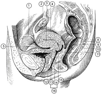
- Supravaginal portion of cervix
Infobox Anatomy
Name = PAGENAME
Latin = portio supravaginalis cervicis
GraySubject = 268
GrayPage = 1259

Caption = 1. Round ligament
2.Uterus
3.Uterine cavity
4.Intestinal surface of Uterus
5.Versical surface (toward bladder)
6.Fundus of uterus
7.Body of uterus
8.Palmate folds of cervical canal
9.Cervical canal
10.Posterior lip
11.Cervical os (external)
12.Isthmus of uterus
13.Supravaginal portion of cervix
14.Vaginal portion of cervix
15.Anterior lip
16.Cervix
Caption2 = Posterior half of uterus and upper part of vagina. (Vaginal portion of cervix visible but not labeled.)
Precursor =
System =
Artery =
Vein =
Nerve =
Lymph =
MeshName =
MeshNumber =
DorlandsPre = p_30
DorlandsSuf = 12660286
The supravaginal portion of the cervix is separated in front from the bladder by fibrous tissue (parametrium ), which extends also on to its sides and lateralward between the layers of thebroad ligaments .The
uterine arteries reach the margins of the cervix in this fibrous tissue, while on either side theureter runs downward and forward in it at a distance of about 2 cm. from the cervix.Posteriorly, the supravaginal cervix is covered by peritoneum, which is prolonged below on to the posterior vaginal wall, when it is reflected on to the
rectum , forming therectouterine excavation .It is in relation with the rectum, from which it may be separated by coils of
small intestine .External links
*
Wikimedia Foundation. 2010.

