- Nasopharyngeal angiofibroma
-
Nasopharyngeal angiofibroma Classification and external resources 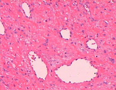
DiseasesDB 32229 MedlinePlus 001572 eMedicine ent/470 Nasopharyngeal angiofibroma (also called juvenile nasopharyngeal angiofibroma)[1][2] is a histologically benign but locally aggressive vascular tumor that grows in the back of the nasal cavity. It most commonly affects adolescent males.[3] Patients with nasopharyngeal angiofibroma usually present with one-sided nasal obstruction and recurrent bleeding.
Contents
Symptomology
- Frequent chronic epistaxis (Nose-Bleeds) or blood-tinged nasal discharge
- Nasal obstruction and rhinorrhea
- Conductive hearing loss from eustachian-tube obstruction
- Diplopia, which occurs secondary to erosion into the cranial cavity and pressure on the optic chiasma
- Rarely anosmia, recurrent otitis media, and eye painDiagnosis
If nasopharyngeal angiofibroma is suspected based on physical exam (a smooth submucosal mass in the posterior nasal cavity), imaging studies such as CT or MRI should be performed. Biopsy can lead to extensive bleeding since the tumor is composed of blood vessels without a muscular coat.
Differential diagnosis
- Antro-choanal polyp (benign neoplasm)
- Rhinosporidiosis (as bleeding point is here too)
- Malignancy - nasopharyngeal carcinoma, lymphoma, plasmacytoma, rhabdomyosarcoma
- Chordoma
- Nasopharyngeal cyst
- Pyogenic granuloma
Treatment
Medical treatment is usually given before surgery to reduce the blood loss - includes the usage of Diethylstilbestrol 2-3 wks before surgery to make the surgery less vascular.
Nasopharyngeal angiofibroma is sometimes treated by surgery.[4] Pre-surgery angiography may allow for embolization, reducing intraoperative blood loss. Hypotensive Anaesthesia is the usual modality followed to reduce blood loss. Patients with tumors that have extended into the cranial cavity or whose tumors can't be safely reached by surgery may receive radiation therapy. Also Radiation therapy helps in case where recurrence is the main problem. Endoscopy has recently been used in patients with tumors of limited extension that have been pre-operatively embolized.
References
- ^ 00021 at CHORUS
- ^ "juvenile nasopharyngeal angiofibroma" at Dorland's Medical Dictionary
- ^ Raphael Rubin; David S. Strayer; Emanuel Rubin (2008). Rubin's Pathology: clinicopathologic foundations of medicine. Lippincott Williams & Wilkins. pp. 1071–. ISBN 9780781795166. http://books.google.com/?id=kD9VZ267wDEC&pg=PA1071. Retrieved 29 June 2010.
- ^ Douglas R, Wormald P (2006). "Endoscopic surgery for juvenile nasopharyngeal angiofibroma: where are the limits?". Curr Opin Otolaryngol Head Neck Surg 14 (1): 1–5. doi:10.1097/01.moo.0000188859.91607.65. PMID 16467630.
Tumors: Mediastinal tumors/Thoracic neoplasm/respiratory neoplasia (C30–C34/D14, 160–163/212.0–212.4) Upper RT Lower RT Tracheal tumorSquamous cell carcinoma · Adenocarcinoma of the lung · Large-cell lung carcinoma · Rhabdoid carcinoma · Sarcomatoid carcinoma · Carcinoid · Salivary gland-like carcinoma of the lung · Adenosquamous carcinoma · Papillary adenocarcinomaNon-carcinomaBy locationPleura Categories:- Head and neck cancer of respiratory tract

Wikimedia Foundation. 2010.