- Color perception test
-
Color perception test Intervention 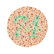
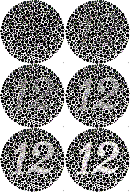
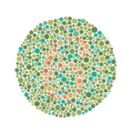
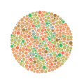
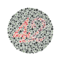
Example of an Ishihara color test plate. The numeral "74" should be clearly visible to viewers with normal color vision. Viewers with dichromat or anomalous trichromat may read it as "21", and viewers with achromat may see nothing.ICD-9-CM 95.06 MeSH D003119 The Ishihara Color Test is an example of a color perception test for red-green color deficiencies. It was named after its designer, Dr. Shinobu Ishihara, a professor at the University of Tokyo, who first published his tests in 1917. [1]
The test consists of a number of colored plates, called Ishihara plates, each of which contains a circle of dots appearing randomized in color and size. Within the pattern are dots which form a number visible to those with normal color vision and invisible, or difficult to see, for those with a red-green color vision defect. The full test consists of 38 plates, but the existence of a deficiency is usually clear after a few plates. Testing the first 24 plates gives a more accurate diagnosis of the severity of the color vision defect.
Common plates include a circle of dots in shades of green and light blues with a figure differentiated in shades of brown, or a circle of dots in shades of red, orange and yellow with a figure in shades of green; the first testing for protanopia and the second for deuteranopia.
Contents
Gallery
See also
References
- ^ S. Ishihara, Tests for colour-blindness (Handaya, Tokyo, Hongo Harukicho, 1917).
External links
- Ishihara color test information
- The Ishihara Color Blindness Test
- Test of color blindness with the indication of the weaknesses in various colours, quantifies green red blue deficiency (ISHIHARA test alternative)
- Flash animated Ishihara color blindness test
Surgery · eye surgery and other procedures (ICD-9-CM V3 08-16+95.0-95.2, ICD-10-PCS 08) Adnexa Lacrimal systemGlobe Radial keratotomy · LASIK · Keratomileusis · Epikeratophakia · Corneal transplantation · Photorefractive keratectomy · Intracorneal ringsExtraocular muscles Medical imaging Fluorescein angiography · Fundus photography · Corneal topography · Optical coherence tomography
Electrodiagnosis: Electrooculography · Electroretinography · ElectronystagmographyEye examination Gonioscopy · Dilated fundus examination · Ocular tonometry · Ophthalmoscopy · Retinoscopy · Color perception test · PerimetryRadiotherapy M: EYE
anat(g/a/p)/phys/devp/prot
noco/cong/tumr, epon
proc, drug(S1A/1E/1F/1L)
Wikimedia Foundation. 2010.

