- Occlusal trauma
-
Not to be confused with Bluetooth.
Occlusal trauma is a dental term that refers to the damage incurred when teeth are left in traumatic occlusion without proper treatment.[1]
When the maxillary and mandibular dental arches approach each together, as they do, for example, during chewing or at rest, the relationship between the opposing teeth is referred to as occlusion. If this occlusal relationship is not balanced properly it may result in pain, tenderness and even mobility of the affected teeth.[1]
When the natural course of trauma, disease and dental treatment alters an individual's occlusion by removing or changing the occlusal (biting) surface of any of the teeth, that individual's teeth will come together, or occlude, differently, and their occlusion will change.[2] When that change is detrimental to the manner in which the teeth occlude, the patient is said to possess a traumatic occlusion.[3] Traumatic occlusion may cause a thickening of the cervical margin of the alveolar bone[4] and widening of the periodontal ligament, although the latter is not pathognomonic for this condition.[5]
Contents
Histologic features associated with occlusal trauma
Microscopically, there will be a number of features that accompany occlusal trauma[6]:
- Hemorrhage
- Necrosis
- Widening of the periodontal ligament, or PDL (also serves as a very common radiographic feature)
- Bone resorption
- Cementum loss and tears
It was concluded that widening of the periodontal ligament was a "functional adaptation to changes in functional requirements".[7]
Clinical signs and symptoms associated with occlusal trauma
Clinically, there are a number of physiologic results that serve as evidence of occlusal trauma[8]:
Primary vs. secondary occlusal trauma
There are two types of occlusal trauma, primary and secondary.
Primary occlusal trauma
Primary occlusal trauma occurs when greater than normal occlusal forces are placed on teeth, as in the case of parafunctional habits, such as bruxism or various chewing or biting habits, including but not limited to those involving fingernails and pencils or pens.
The associated excessive forces can be grouped into three categories. Excesses of[9]:
- Duration
- Frequency and
- Magnitude
Primary occlusal trauma will occur when there is a normal periodontal attachment apparatus and, thus, no periodontal disease.[10]
Secondary occlusal trauma
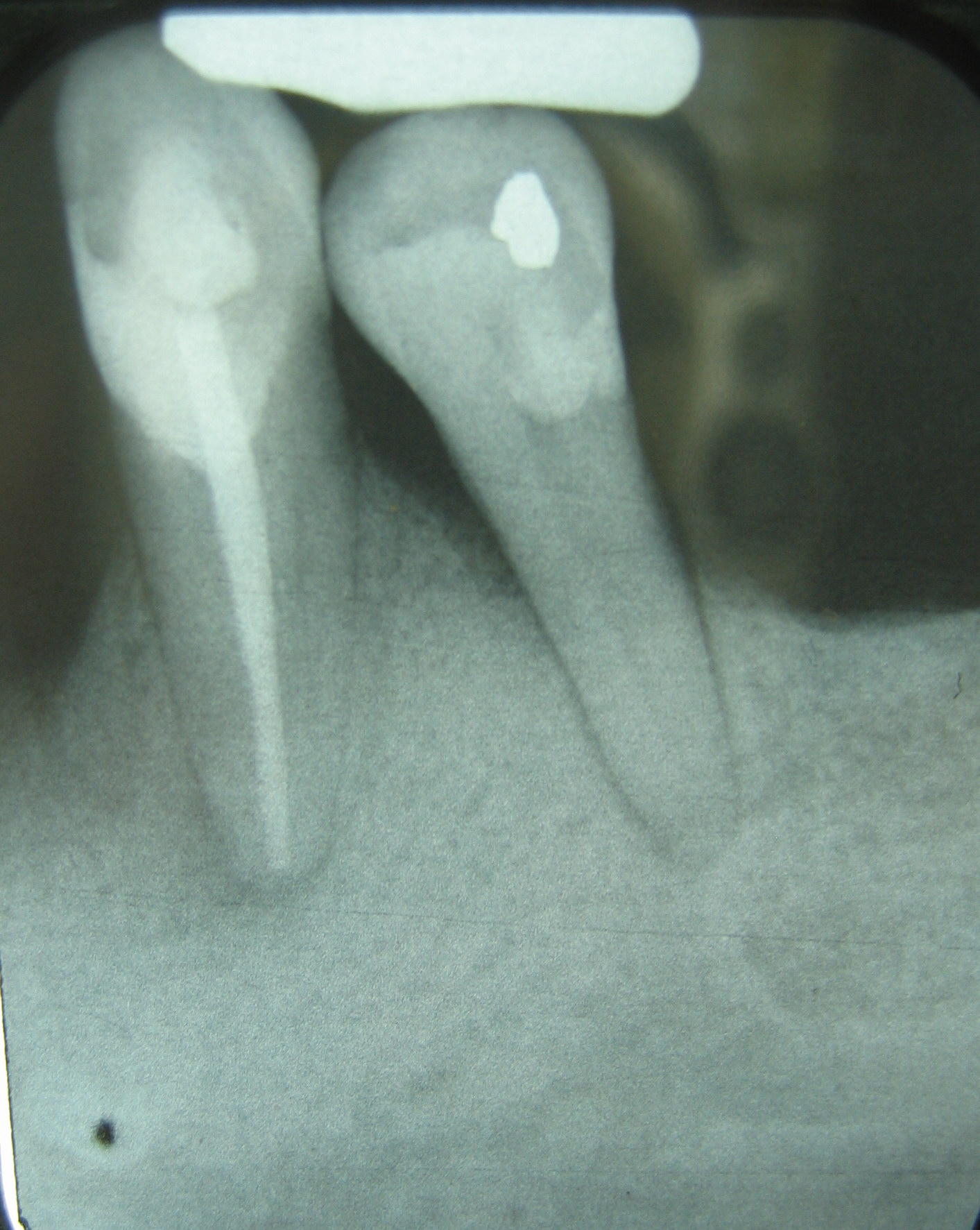
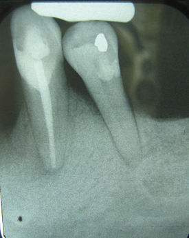 An example of secondary occlusal trauma. This X-ray film displays two lone-standing mandibular teeth, #21 and #22, or the lower left first premolar and canine. As the remnants of a once full complement of 16 lower teeth, these two teeth have been alone in opposing the forces associated with mastication for some time, as can be evidenced by the widened PDL surrounding the premolar Because this trauma is occurring on teeth that have 30-50% bone loss, this would be classified as secondary oclcusal trauma.
Secondary occlusal trauma occurs when normal occlusal forces are placed on teeth with compromised periodontal attachment, thus contributing harm to an already damaged system. As stated, secondary occlusal trauma occurs when there is a compromised periodontal attachment and, thus, a pre-existing periodontal condition.[10]
Etiology and treatment
Teeth are constantly subject to both horizontal and vertical occlusal forces. With the center of rotation of the tooth acting as a fulcrum, the surface of bone adjacent to the pressured side of the tooth will undergo resorption and disappear, while the surface of bone adjacent to the tensioned side of the tooth will undergo apposition and increase in volume.[11]
In both primary and secondary occlusal trauma, tooth mobility might develop over time, with it occurring earlier and being more prevalent in secondary occlusal trauma. To treat mobility due to occlusal trauma, whether it be primary or secondary, the affected teeth are splinted together and to the adjacent teeth so as to eliminate their mobility.
In primary occlusal trauma, the etiology, or cause, of the mobility was the excessive force being applied to a tooth with a normal attachment apparatus, otherwise known as a periodontally-uninvolved tooth. The approach should be to eliminate the etiology of the pain and mobility by determining the causes and removing them; the mobile tooth or teeth will soon cease exhibiting mobility. This could involve removing a high spot on a recently restored tooth, or even a high spot on a non-recently restored tooth that perhaps moved into hyperocclusion. It could also involved altering ones parafunctional habits, such as refraining from chewing on pens or biting one's fingernails. For a bruxer, treatment of the patient's primary occlusal trauma could involve selective grinding of certain interarch tooth contacts or perhaps employing a nightguard to protect the teeth from the greater than normal occlusal forces of the patient's parafunctional habit. For someone who is missing enough teeth in non-strategic positions so that the remaining teeth are forced to endure a greater per square inch occlusal force, treatment might include restoration with either a removable prosthesis or implant-supported crown or bridge.
In secondary occlusal trauma, simply removing the "high spots" or selective grinding of the teeth will not eliminate the problem, because the teeth are already periodontally involved. After splinting the teeth to eliminate the mobility, the etiology of the mobility (in other words, the loss of clinical attachment and bone) must be managed; this is achieved through surgical periodontal procedures such as soft tissue and bone grafts, as well as restoration of edentulous areas. As with primary occlusal trauma, treatment may include either a removable prosthesis or implant-supported crown or bridge.
References
- ^ a b Bibb, CA: Occlusal Evaluation and Therapy in the Management of Periodontal Disease. In Newman, MG; Takei, HH; Carranza, FA; editors: Carranza’s Clinical Periodontology, 9th Edition. Philadelphia: W.B. Saunders Company, 2002. pages 698-701.
- ^ Hinrichs, JE: Occlusal The Role of Dental Calculus and Other Predisposing Factors. In Newman, MG; Takei, HH; Carranza, FA; editors: Carranza’s Clinical Periodontology, 9th Edition. Philadelphia: W.B. Saunders Company, 2002. page 192.
- ^ traumatogenic occlusion - definition of traumatogenic occlusion in the Medical dictionary - by the Free Online Medical Dictionary, Thesaurus and Encyclopedia
- ^ Carranza, FA: Bone Loss and Patterns of Bone Destructions. In Newman, MG; Takei, HH; Carranza, FA; editors: Carranza’s Clinical Periodontology, 9th Edition. Philadelphia: W.B. Saunders Company, 2002. page 362.
- ^ Trauma from Occlusion Handout, Dr. Michael Deasy, Department of Periodontics, NJDS 2007. page 5
- ^ Trauma from Occlusion Handout, Dr. Michael Deasy, Department of Periodontics, NJDS 2007. page 7
- ^ Wentz et al. J Perio, 1958
- ^ Trauma from Occlusion Handout, Dr. Michael Deasy, Department of Periodontics, NJDS 2007. page 12
- ^ Trauma from Occlusion Handout, Dr. Michael Deasy, Department of Periodontics, NJDS 2007. page 14
- ^ a b Carranza, FA; Bernard, GW: The Tooth-Supporting Structures. In Newman, MG; Takei, HH; Carranza, FA; editors: Carranza’s Clinical Periodontology, 9th Edition. Philadelphia: W.B. Saunders Company, 2002. page 53.
- ^ Trauma from Occlusion Handout, Dr. Michael Deasy, Department of Periodontics, NJDS 2007. page 4
Periodontology Tissues of the periodontium
and their physiologic entitiesAlveolar bone · Biologic width · Bundle bone · Cementum · Free gingival margin · Gingiva · Gingival fibers · Gingival sulcus · Junctional epithelium · Mucogingival junction · Periodontal ligament · Sulcular epithelium · StipplingDiagnoses Chronic periodontitis · Localized aggressive periodontitis · Generalized aggressive periodontitis · Periodontitis as a manifestation of systemic disease · Necrotizing periodontal diseases · Abscesses of the periodontium · Combined periodontic-endodontic lesionsPathogenesis A. actinomycetemcomitans · Capnocytophaga sp. · F. nucleatum · P. gingivalis · P. intermedia · T. forsythia · T. denticolaPathologic entities Calculus · Clinical attachment loss · Edentulism · Fremitus · Furcation defect · Gingival enlargement · Gingival pocket · Gingivitis · Horizontal bony defect · Linear gingival erythema · Occlusal trauma · Periodontal pocket · Periodontal disease · Periodontitis · Plaque · Recession · Vertical bony defectDiagnosis, treatment planning,
prevention and
chemotherapeutic agentsBrushing · Bleeding on probing · Chlorhexidine gluconate · Enamel matrix derivative · Flossing · Hydrogen peroxide · Mouthwash · Oral hygiene · Tetracycline · TriclosanPeriodontal armamentarium Conventional therapy Surgical therapy and
periodontal surgeryApically positioned flap · Bone graft · Coronally positioned flap · Crown lengthening · Open flap debridement · Free gingival graft · Gingivectomy · Guided bone regeneration · Guided tissue regeneration · Implant Placement · Lateral pedicle graft · Pocket reduction surgery · Sinus lift · Subepithelial connective tissue graftImportant personalities Per-Ingvar Brånemark · Jan Lindhe · Preston D. Miller · Willoughby D. Miller · Carl E. Misch · John Mankey Riggs · Jørgen Slots · Dennis P. Tarnow · Hom-Lay Wang · James Leon Williams · W. J. YoungerOther specialties Endodontology · Orthodontology · ProsthodontologyCategories:- Acquired tooth disorders
- Periodontology
Wikimedia Foundation. 2010.