- Great auricular nerve
-
Nerve: Great auricular nerve 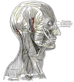
The nerves of the scalp, face, and side of neck. (Great auricular visible below ear.) 
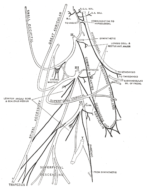
Plan of the cervical plexus. (Great auricular labeled at top center.) Latin nervus auricularis magnus Gray's subject #210 926 Innervates Cutaneous innervation of the inferior part of the auricle and the parotid region of the face. From Cervical plexus (C2-C3) The great auricular nerve originates from the cervical plexus, composed of branches of spinal nerves C2 and C3. It provides sensory innervation for the skin over parotid gland and mastoid process, and both surfaces of the outer ear.
Contents
Terminology
Although this nerve is frequently referred to as the "greater" auricular nerve, this is not the proper nomenclature since there is no "lesser" auricular nerve. Great refers to the distinction between it and the Auriculotemporal nerve, which is the less influential of the two.
Path
It is the largest of the ascending branches. It arises from the second and third cervical nerves, winds around the posterior border of the Sternocleidomastoideus, and, after perforating the deep fascia, ascends upon that muscle beneath the Platysma to the parotid gland, where it divides into an anterior and a posterior branch.
Branches
- The anterior branch (ramus anterior; facial branch) is distributed to the skin of the face over the parotid gland, and communicates in the substance of the gland with the facial nerve.
- The posterior branch (ramus posterior; mastoid branch) supplies the skin over the mastoid process and on the back of the auricula, except at its upper part; a filament pierces the auricula to reach its lateral surface, where it is distributed to the lobule and lower part of the concha. The posterior branch communicates with the smaller occipital, the auricular branch of the vagus, and the posterior auricular branch of the facial.
Additional images
-

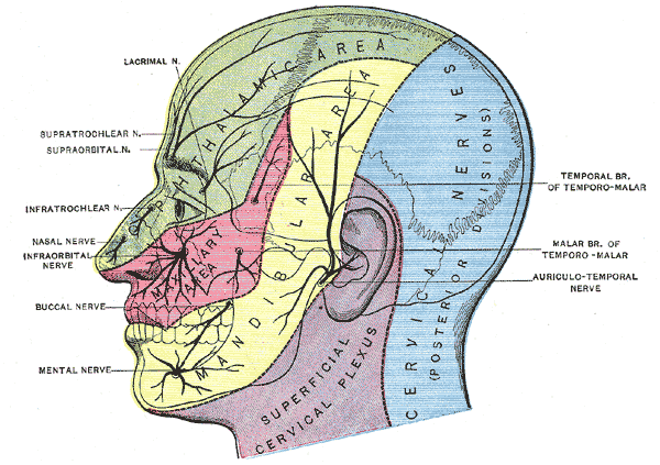
Dermatome distribution of the trigeminal nerve
-
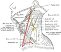
Side of neck, showing chief surface markings.
External links
- Diagram at aapmr.org
- SUNY Figs 25:03-03 - "Diagram of the cervical plexus."
This article was originally based on an entry from a public domain edition of Gray's Anatomy. As such, some of the information contained within it may be outdated.
Nerves of head and neck: the cervical plexus (C1–C4) (TA A14.2.02, GA 9.926) superficial deep Categories:- Nerves of the head and neck
- Neuroanatomy stubs


Wikimedia Foundation. 2010.