- Phytoplasma
-
Phytoplasma 
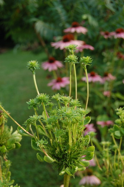
Phyllody induced by phytoplasma infection on a coneflower (Echinacea purpurea). Scientific classification Division: Firmicutes Class: Mollicutes Order: Acholeplasmatales Family: Acholeplasmataceae Genus: Candidatus Phytoplasma Species "Ca. Phytoplasma allocasuarinae"
"Ca. Phytoplasma asteris"
"Ca. Phytoplasma aurantifolia"
"Ca. Phytoplasma australiense"
"Ca. Phytoplasma brasiliense"
"Ca. Phytoplasma castaneae"
"Ca. Phytoplasma cocostanzaniae"
"Ca. Phytoplasma cocosnigeriae"
"Ca. Phytoplasma cynodontis"
"Ca. Phytoplasma fraxini"
"Ca. Phytoplasma japonicum"
"Ca. Phytoplasma luffae"
"Ca. Phytoplasma mali"
"Ca. Phytoplasma oryzae"
"Ca. Phytoplasma palmae"
"Ca. Phytoplasma phoenicium"
"Ca. Phytoplasma pruni"
"Ca. Phytoplasma prunorum"
"Ca. Phytoplasma pyri"
"Ca. Phytoplasma rhamni"
"Ca. Phytoplasma solani"
"Ca. Phytoplasma spartii"
"Ca. Phytoplasma trifolii"
"Ca. Phytoplasma ulmi"
"Ca. Phytoplasma vitis"
"Ca. Phytoplasma ziziphi"Phytoplasma are specialised bacteria that are obligate parasites of plant phloem tissue and transmitting insects (vectors). They were first discovered by scientists in 1967 and were named mycoplasma-like organisms or MLOs.[1] They cannot be cultured in vitro in cell-free media. They are characterised by their lack of a cell wall, a pleiomorphic or filamentous shape, normally with a diameter less than 1 micrometer, and their very small genomes.
Phytoplasmas are pathogens of important plants, including coconut, sugarcane, sandal wood, causing a wide variety of symptoms that range from mild yellowing to death of infected plants. They are most prevalent in tropical and sub-tropical regions of the world. Phytoplasmas require a vector to be transmitted from plant to plant, and this normally takes the form of sap sucking insects such as leaf hoppers in which they are also able to replicate.
Contents
History
There are references to diseases now known to be caused by phytoplasmas as far back as 1603 for Mulberry dwarf disease in Japan.[2] Such diseases were originally thought to be caused by viruses, which, like phytoplasmas, require insect vectors, cannot be cultured, and have some symptom similarity.[3] In 1967 phytoplasmas were discovered in ultrathin sections of plant phloem tissue and named mycoplasma-like organisms (MLOs), because they physically resembled mycoplasmas[1] The organisms were renamed phytoplasmas in 1994 at the 10th congress of the International Organization of Mycoplasmology.[3]
Morphology
Being mollicutes, phytoplasmas lack cell walls and instead are bound by a triple layered membrane.[4] The cell membranes of all phytoplasmas studied so far usually contain a single immunodominant protein (of unknown function) that makes up the majority of the protein content of the cell membrane.[5] The typical phytoplasma exhibits a pleiomorphic or filamentous shape and is less than 1 micrometer in diameter. Like other prokaryotes, DNA is free in the cytoplasm.
Symptoms
A common symptom caused by phytoplasma infection is phyllody, the production of leaf-like structures in place of flowers. Evidence suggests that the phytoplasma downregulates a gene involved in petal formation (AP3 and its orthologues) and genes involved in the maintenance of the apical meristem (Wus and CLV1).[6] Other symptoms, such as the yellowing of leaves, are thought to be caused by the phytoplasma's presence in the phloem, affecting its function and changing the transport of carbohydrates.[7]
Phytoplasma infected plants may also suffer from virescence, the development of green flowers due to the loss of pigment in the petal cells.[8] Sometimes sterility of the flowers is also seen.
Many phytoplasma infected plants gain a bushy or witch's broom appearance due to changes in normal growth patterns caused by the infection. Most plants show apical dominance, but phytoplasma infection can cause the proliferation of auxiliary (side) shoots and an increase in size of the internodes.[8] Such symptoms are actually useful in the commercial production of poinsettia. The infection produces more axillary shoots, which enables production of poinsettia plants that have more than one flower.[9]
Phytoplasmas may cause many other symptoms that are induced because of the stress placed on the plant by infection rather than specific pathogenicity of the phytoplasma. Photosynthesis, especially photosystem II, is inhibited in many phytoplasma infected plants.[4] Phytoplasma infected plants often show yellowing which is caused by the breakdown of chlorophyll, whose biosynthesis is also inhibited.[4]
Transmission
Movement between plants
The phytoplasmas are mainly spread by insects of the families Cicadellidea (leafhoppers), Fulgoridea (planthoppers) and Psyllidae (jumping plant lice) ,[10] which feed on the phloem tissues of infected plants, picking up the phytoplasmas and transmitting them to the next plant they feed on. For this reason the host range of phytoplasmas is strongly dependent upon its insect vector. Phytoplasmas contain a major antigenic protein that makes up the majority of their cell surface proteins. This protein has been shown to interact with insect microfilament complexes and is believed to be the determining factor in insect-phytoplasma interaction.[11] Phytoplasmas may overwinter in insect vectors or perennial plants. Phytoplasmas can have varying effects on their insect hosts; examples of both reduced and increased fitness have been seen.[12]
Phytoplasmas enter the insect's body through the stylet, move through the intestine, and are then absorbed into the haemolymph.[12] From here they proceed to colonise the salivary glands, a process that can take up to three weeks.[12] Once established, phytoplasmas will be found in most major organs of an infected insect host. The time between being taken up by the insect and reaching an infectious titre in the salivary glands is called the latency period.[12]
Phytoplasmas can also be spread via dodders cascutaceae[13] or vegetative propagation such as the grafting of a piece of infected plant onto a healthy plant.
Movement within plants
Phytoplasmas are able to move within the phloem from source to sink, and they are able to pass through sieve tube elements. But since they spread more slowly than solutes, for this and other reasons, movement by passive translocation is not supported.[14]
Detection and Diagnosis
Before molecular techniques were developed, the diagnosis of phytoplasma diseases was difficult because they could not be cultured. Thus classical diagnostic techniques, such as observation of symptoms, were used. Ultrathin sections of the phloem tissue from suspected phytoplasma infected plants would also be examined for their presence.[1] Treating infected plants with antibiotics such as tetracycline to see if this cured the plant was another diagnostic technique employed.
Molecular diagnostic techniques for the detection of phytoplasma began to emerge in the 1980s and included ELISA based methods. In the early 1990s, PCR-based methods were developed that were far more sensitive than those that used ELISA, and RFLP analysis allowed the accurate identification of different strains and species of phytoplasma.[15]
More recently, techniques have been developed that allow for assessment of the level of infection. Both QPCR and bioimaging have been shown to be effective methods of quantifying the titre of phytoplasmas within the plant.[14]
Control
Phytoplasmas are normally controlled by the breeding and planting of disease resistance varieties of crops (believed to the most economically viable option) and by the control of the insect vector.[8]
Tissue culture can be used to produce clones of phytoplasma infected plants that are healthy. The chances of gaining healthy plants in this manner can be enhanced by the use of cryotherapy, freezing the plant samples in liquid nitrogen, before using them for tissue culture.[16]
Work has also been carried out investigating the effectiveness of plantibodies targeted against phytoplasmas.[17]
Tetracyclines are bacteriostatic to phytoplasmas, that is they inhibit their growth.[18] However, without continuous use of the antibiotic, disease symptoms will reappear. Thus, tetracycline is not a viable control agent in agriculture, but it is used to protect ornamental coconut trees.[19]
Genetics
The genomes of three phytoplasmas have been sequenced: Aster Yellows Witches Broom,[20] Onion Yellows (Ca. Phytoplasma asteris)[21] and Ca. Phytoplasma australiense[22] Phytoplasmas have very small genomes, which also have extremely low levels of the nucleotides G and C, sometimes as little as 23% which is thought to be the threshold for a viable genome.[23] In fact Bermuda grass white leaf phytoplasma has a genome size of just 530Kb, one of the smallest known genomes of living organisms.[24] Larger phytoplasma genomes are around 1350 Kb. The small genome size associated with phytoplasmas is due to their being the product of reductive evolution from Bacillus/Clostridium ancestors. They have lost 75% or more of their original genes, and this is why they can no longer survive outside of insects or plant phloem. Some phytoplasmas contain extrachromosomal DNA such as plasmids.[25]
Despite their very small genomes, many predicted genes are present in multiple copies. Phytoplasmas lack many genes for standard metabolic functions and have no functioning homologous recombination pathways, but do have a sec transport pathway.[20] Many phytoplasmas contain 2 rRNA operons. Unlike the rest of the Mollicutes, the triplet code of UGA is used as a stop codon in phytoplasmas, rather than to code for tryptophan.[26]
Phytoplasma genomes contain large numbers of transposon genes and insertion sequences. They also contain a unique family of repetitive extragenic palindromes (REPs) called PhREPS whose role is unknown though it is theorised that the stem loop structures the PhREPS are capable of forming may play a role in transcription termination or genome stability.[27]
Taxonomy
Phytoplasmas are mollicutes and within this group belong to the monophyletic order Acholeplasmatales.[8] In 1992 the Subcommittee on the Taxonomy of Mollicutes proposed the use of the name Phytoplasma in place of the use of the term MLO (Mycoplasma-like organism) "for reference to the phytopathogenic mollicutes".[28] In 2004 the genus name Phytoplasma was adopted and is currently at Candidatus status[29] which is used for bacteria that can not be cultured.[30] It's taxonomy is complicated by the fact that it can not be cultured and thus methods normally used for classification of prokaryotes are not possible.[8] Phytoplasma taxonomic groups are based on differences in the fragment sizes produced by the restriction digest of the 16S rRNA gene sequence (Called RFLP) or by comparison of DNA sequences from the 16s/23s spacer regions.[31] There is some disagreement over how many taxonomic groups the phytoplasmas fall into, recent work involving computer simulated restriction digests of the 16Sr gene suggest there may be up to 28 groups[32] where as other papers argue for less groups, but more sub-groups. Each group includes at least one Ca. Phytoplasma species, characterised by distinctive biological, phytopathological and genetic properties. The table below summaries some of the major taxonomic groups and the candidatus species that belong in them.
16Sr Group Group Name Species Reference 16SrI Aster yellows Ca. Phytoplasma asteris
Ca. Phytoplasma japonicum16SrII Peanut witch's broom Ca. Phytoplasma aurantifolia 16SrIII X-disease Ca. Phytoplasma pruni 16SrIV Coconut lethal yellowing Ca. Phytoplasma palmae
Ca. Phytoplasma castaneae
Ca. Phytoplasma cocosnigeriae16SrV Elm yellows Ca. Phytoplasma ziziphi
Ca. Phytoplasma vitis
Ca. Phytoplasma ulmi16SrVI Clover proliferation Ca. Phytoplasma trifolii 16SrVII Ash yellows Ca. Phytoplasma fraxini 16SrVIII Luffa witch's-broom Ca. Phytoplasma luffae 16SrIX Pidgeon pea witch's broom Ca. Phytoplasma phoenicium 16SrX Apple proliferation Ca. Phytoplasma mali
Ca. Phytoplasma pyri
Ca. Phytoplasma prunorum
Ca. Phytoplasma spartii
Ca. Phytoplasma rhamnii
Ca. Phytoplasma allocasuarinae16SrXI Rice Yellow Dwarf
Sugarcane Grassy Shoot Disease (SCGS)Ca. Phytoplasma oryzae [33] 16SrXII Stolbur Ca. Phytoplasma solani
Ca. Phytoplasma australiense16SrXIII Mexican periwinkle virescence Undefined 16SrXIV Bermuda white leaf Ca. Phytoplasma cynodontis 16SrXV Hibiscus witch's-broom Ca. Phytoplasma brasiliense Gallery
-
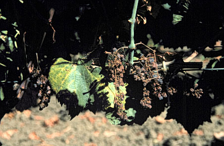
A grape vine with phytoplasma disease
-

Coconut palms dying of lethal yellowing disease
-
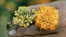
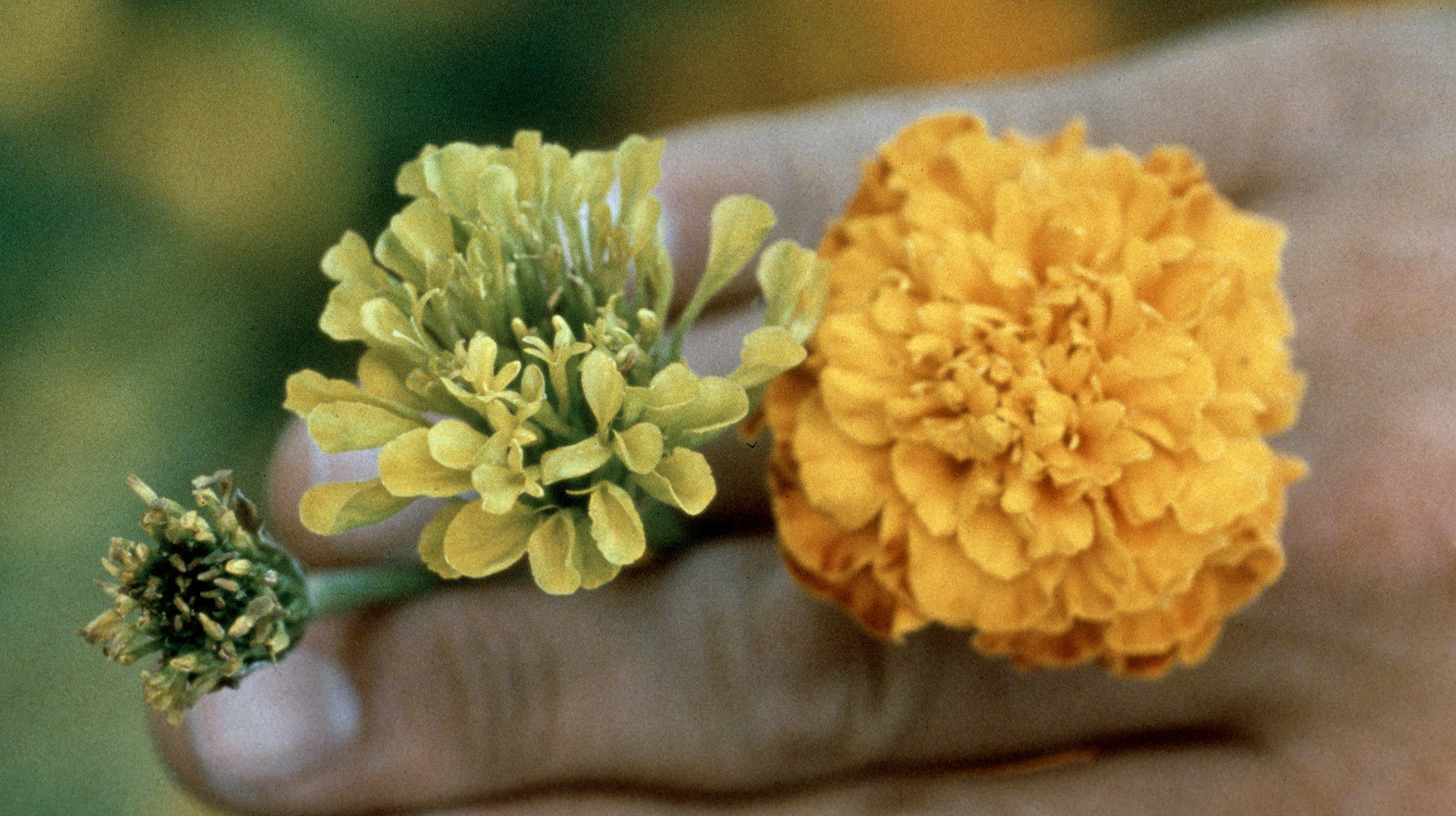
Symptoms of Aster yellows on marigold
-

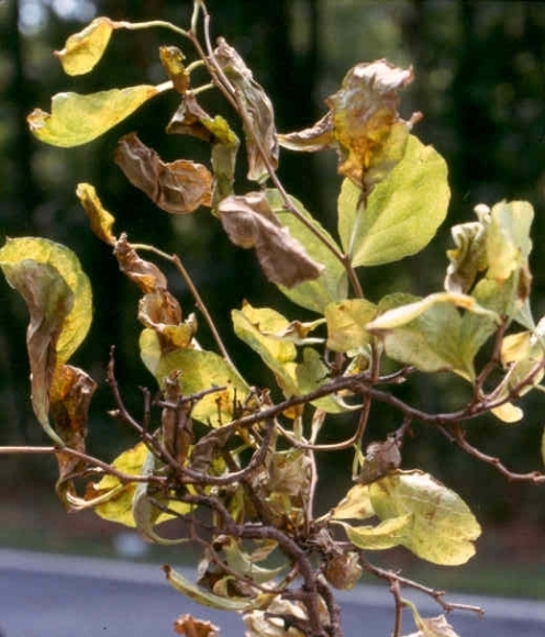
Symptoms of elm phloem necrosis phytoplasma
-
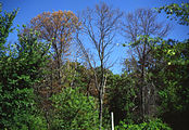
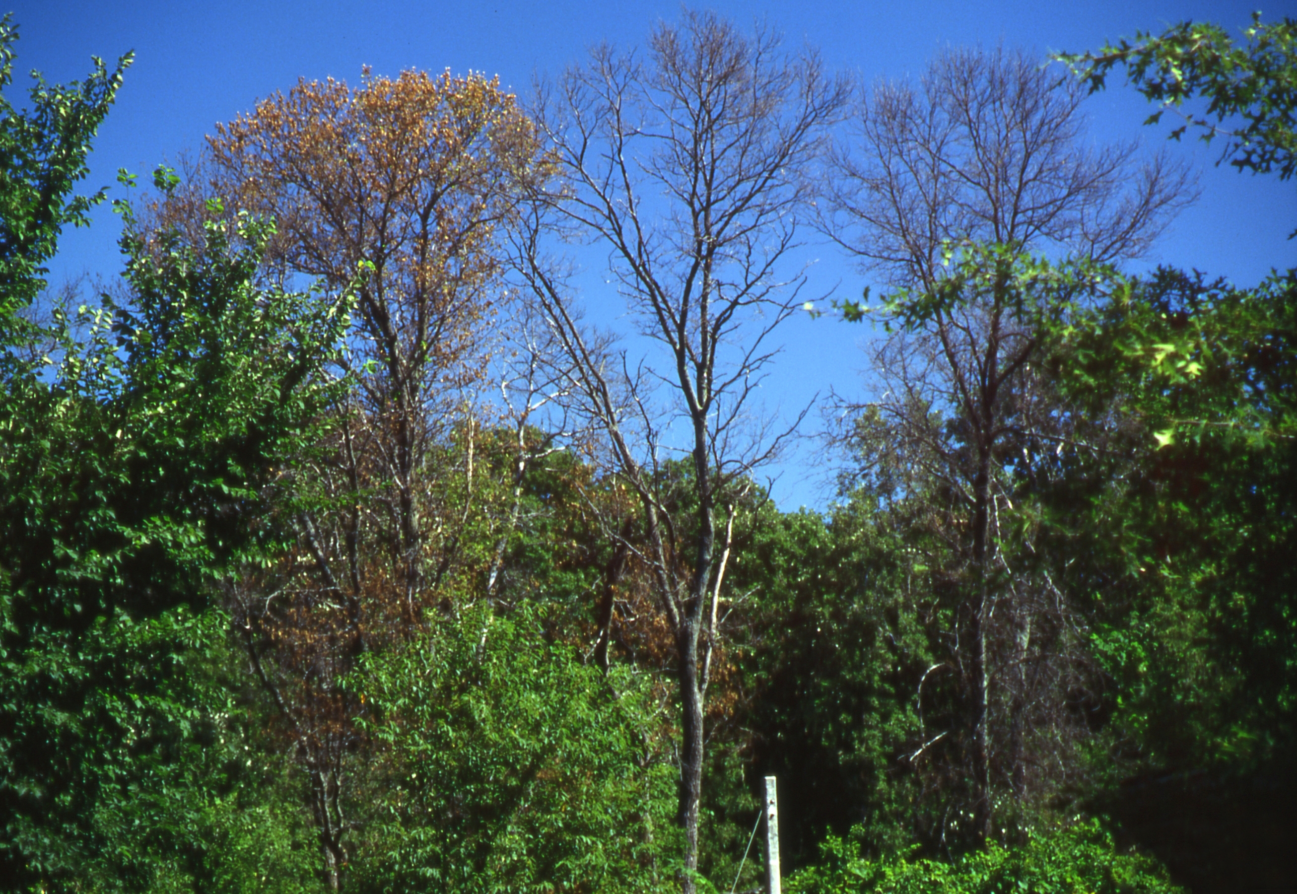
Trees dying of Ash yellows phytoplasma
-
Phyllody caused by phytoplasma infection on Cosmos spp.
-

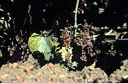
Symptoms of sweet potato little leaf phytoplasma on Catharanthus roseus
-

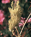
Phyllody of goldenrod
-

A palm tree dying of lethal yellowing phytoplasma
See also
- Mollicutes
- Mycoplasma
- Polymerase Chain Reaction or PCR
- ELISA
- Sugarcane Grassy Shoot Disease (SCGS)
References
- ^ a b c Doi, Y; Teranaka M, Yora, K and Asuyama, H (1967). "Mycoplasma or PLT-group-like organisms found in the phloem elements of plants infected with mulberry dwarf, potato witches' broom, aster yellows or paulownia witches' broom". Annals of the Phytopathological Society of Japan 33: 259–266.
- ^ Okuda, S (1972). "Occurrence of diseases caused by mycoplasma-like organisms in Japan". Plant Protection 26: 180–183.
- ^ a b Hogenhout, SA; Oshima K, Ammar E-D, Kakizawa S, Kingdom HN and Namba S (2008). "Phytoplasmas: bacteria that manipulate plants and insects". Molecular Plant Pathology 9 (4): 403–423. doi:10.1111/j.1364-3703.2008.00472.x. PMID 18705857. http://www3.interscience.wiley.com/journal/119879040/abstract. Retrieved 2008-07-04.
- ^ a b c Bertamini, M; Grando M. S and Nedunchezhian N (2004). "Effects of Phytoplasma Infection on Pigments, Chlorophyll-Protein Complex and Photosynthetic Activities in Field Grown Apple Leaves". Biologia Plantarum (Springer Netherlands) 47 (2): 237–242. doi:10.1006/pmpp.2003.0450. http://www.springerlink.com/content/xn2k8h287846611p/.
- ^ Berg, M; Davis DL, Clark MF, Vetten HJ, Maier G, Marcone C and Seemuller E (1999). "Isolation of the gene encoding an immunodominant membrane protein of the apple proliferation phytoplasma, and expression and characterization of the gene product". Microbiology (Society for General Microbiology) 145: 1939–1943. PMID 10463160. http://mic.sgmjournals.org/cgi/content/abstract/145/8/1937.
- ^ Pracros, P; Renaudin J, Eveillard S, Mouras A and Hernould M (2006). "Tomato Flower Abnormalities Induced by Stolbur Phytoplasma Infection Are Associated with Changes of Expression of Floral Development Genes". Molecular Plant Microbe Interactions (APS Press) 19 (1): 62–68. doi:10.1094/MPMI-19-0062. PMID 16404954. http://apsjournals.apsnet.org/doi/abs/10.1094/MPMI-19-0062.
- ^ Muast BE, Espadas F, Talavera C, Aguilar M, Santamaría JM, Oropeza C (2003). "Changes in carbohydrate metabolism in coconut palms infected with the lethal yellowing phytoplasma". Phytopathology 93 (8): 976–981. doi:10.1094/PHYTO.2003.93.8.976. PMID 18943864.
- ^ a b c d e Lee, IM; Davis RE and Gundersen-Rindal DE (2000). "Phytoplasma: Phytopathogenic Mollicutes". Annual Review of Microbiology (Annual Reviews) 54: 221–255. doi:10.1146/annurev.micro.54.1.221. PMID 11018129. http://arjournals.annualreviews.org/doi/abs/10.1146%2Fannurev.micro.54.1.221.
- ^ Lee, IM; Klopmeyer M, Bartoszyk IM, Gundersen-Rindal DE, Chou TS, Thomson KL and Eisenreich R (1997). "Phytoplasma induced free-branching in commercial poinsettia cultivars". Nature Biotechnology (Nature Publishing Group) 15 (2): 178–182. doi:10.1038/nbt0297-178. PMID 9035146. http://www.nature.com/nbt/journal/v15/n2/abs/nbt0297-178.html.
- ^ Weintraub, Phyllis G.; Beanland, LeAnn (2006). "Insect vectors of phytoplasmas". Annual Review of Entomology 51: 91–111. doi:10.1146/annurev.ento.51.110104.151039. PMID 16332205. http://arjournals.annualreviews.org/doi/abs/10.1146/annurev.ento.51.110104.151039.
- ^ Suzuki et al. (2006). "Interactions between a membrane protein of a pathogen and insect microfilament complex determines insect vector specificity". Proceedings of the National Academy of Sciences 103: 4252–4257. doi:10.1073/pnas.0508668103.
- ^ a b c d Christensen N, Axelsen K, Nicolaisen M, Schulz A (2005). "Phytoplasmas and their interactions with their hosts". Trends in Plant Sciences 10: 526–535. doi:10.1016/j.tplants.2005.09.008.
- ^ Carraro, L.; Loi, N.; Favali, M. A.; Favali, M. A. (1991). "Transmission characteristics of the clover phyllody agent by dodder". J. Phytopathol 133: 15–22. doi:10.1111/j.1439-0434.1991.tb00132.x.
- ^ a b Christensen NM, Nicolaisen M, Hansen M, Schulz A (2004). "Distribution of phytoplasmas in infected plants as revealed by real time PCR and bioimaging". Molecular plant microbe interactions 17 (11): 1175–1184. doi:10.1094/MPMI.2004.17.11.1175. PMID 15553243.
- ^ Chen et al. (1992) Detection and identification of plant and insect mollicutes. In The Mycoplasmas, editor RF Whitcomb and JG Tully 5: 393-424
- ^ Wang et al. (2007) Effective elimination of sweet potato little lead by cryotherapy of shoot tips. Plant Pathology online early edition.
- ^ Chen, Y. D.; Chen, T. A. (1998). "Expression of engineered antibodies in plants: A possible tool for spiroplasma and phytoplasma disease control". Phytopathology 88 (12): 1367–1371. doi:10.1094/PHYTO.1998.88.12.1367. PMID 18944841.
- ^ Davies R. E., Whitcomb R. F., Steere R. L. (1968). "Remission of aster yellows disease by antibiotics". Science 161 (3843): 793–794. doi:10.1126/science.161.3843.793.
- ^ Drug for Humans Checks Palm Trees Disease. New York Times, July 19 1983
- ^ a b Bai X, Zhang J, Ewing A, Miller SA, Jancso Radek A, Shevchenko DV, Tsukerman K, Walunas T et al. (2006). "Living with genome instability: the adaption of phytoplasmas to diverse environments of their insect and plant hosts". Journal of Bacteriology 188 (10): 3682–3696. doi:10.1128/JB.188.10.3682-3696.2006. PMC 1482866. PMID 16672622. http://www.pubmedcentral.nih.gov/articlerender.fcgi?tool=pmcentrez&artid=1482866.
- ^ Oshima K, Kakizawa S, Nishigawa H, Jung HY, Wei W, Suzuki S, Arashida R, Nakata D et al. (2004). "Reductive evolution suggested from the complete genome sequence of a plant-pathogenic phytoplasma". Nature Genetics 36 (1): 27–29. doi:10.1038/ng1277. PMID 14661021.
- ^ Tran-Nguyen, LT; Kube, M; Schneider, B; Reinhardt, R; Gibb, KS (2008). "Comparative genome analysis of "Candidatus Phytoplasma australiense" (subgroup tuf-Australia I; rp-A) and "Ca. Phytoplasma asteris" Strains OY-M and AY-WB.". Journal of bacteriology 190 (11): 3979–91. doi:10.1128/JB.01301-07. PMC 2395047. PMID 18359806. http://www.pubmedcentral.nih.gov/articlerender.fcgi?tool=pmcentrez&artid=2395047.
- ^ Dikinson, M. Molecular Plant Pathology (2003) BIOS Scientific Publishers
- ^ Marcone, C; Neimark H, Ragozzino A, Lauer U and Seemüller E (1999). "Chromosome Sizes of Phytoplasmas Composing Major Phylogenetic Groups and Subgroups". Phytopathology (APS Press) 89 (9): 805–810. doi:10.1094/PHYTO.1999.89.9.805. PMID 18944709. http://apsjournals.apsnet.org/doi/abs/10.1094/PHYTO.1999.89.9.805.
- ^ Nishigawa et al. (2003). "Complete set of extrachromosomal DNAs from three pathogenic lines of onion yellows phytoplasma and use of PCR to differentiate each line". Journal of General Plant Pathology 69: 194–198. doi:1007/s10327-003-0035-1.
- ^ Razin S, Yogev D, Naot Y (1998). "Molecular biology and pathogenicity of mycoplasmas". Microbiology Molecular Biology Review 62 (4): 1094–1156. PMC 98941. PMID 9841667. http://www.pubmedcentral.nih.gov/articlerender.fcgi?tool=pmcentrez&artid=98941.
- ^ Jomantiene R, Davis RE (2006). "Clusters of diverse genes existing as multiple, sequence variable mosaics in a phytoplasma genomes". FEMS Microbiology letters 255 (1): 59–65. doi:10.1111/j.1574-6968.2005.00057.x. PMID 16436062.
- ^ Subcommittee on the Taxonomy of Mollicutes. Minutes of the Interim Meetings, 1 and 2 August, 1992, Ames, Iowa Int. J. of Syst. Bact. April 1993, p. 394-397; Vol. 43, No. 2 (see minutes 10 and 25)
- ^ The IRPCM Phytoplasma/Spiroplasma Working Team - Phytoplasma taxonomy group: Candidatus Phytoplasma, a taxon for the wall-less, non-helical prokaryotes that colonize plant phloem and insects. Int. J. Syst. Evol. Microbiol., 2004, 54, 1243-1255.[1]
- ^ Murry, Syst Bacteriol et al. (1995). '. 45. pp. 186–187.
- ^ Hodgetts et al. (2007). "Taxonomic groupings based on the analysis on the 16s/23s spacer regions which shows greater variation than the normally used 16srRNA gene results in classification similar to that derived from 16s rRNA data but with more detailed subdivisions". Plant Pathology 56: 357–365.
- ^ Wei W, Davis RE, Lee IM, Zhao Y (2007). "Computer-simulated RFLP analysis of 16S rRNA genes: identification of ten new phytoplasma groups". International journal of systematic and evolutionary microbiology 57 (Pt 8): 1855–1867. doi:10.1099/ijs.0.65000-0. PMID 17684271.
- ^ a b Nasare, Kanchan; Yadav, Amit; Singh, Anil K.; Shivasharanappa, K. B.; Nerkar, Y. S.; Reddy, V. S. (2007). "Molecular and symptom analysis reveal the presence of new phytoplasmas associated with sugarcane grassy shoot disease in India". Plant Disease 91: 1413. doi:10.1094/PDIS-91-11-1413.
External links
- First International Phytoplasmologist Working Group Meeting published in Vol. 60-2 2007 of Bulletin of Insectology
- Photo gallery about plants infected of phytoplasma
- Phytoplasma Resource and phytoplasma classification database
- Ohio State University publishes an informative site on this topic.
- First Internet Conference of Phytopathogenic Mollicutes includes several interesting articles on this topic.
- Phytoplasma Genome Projects.
- The Centre for Information on Coconut Lethal Yellowing (CICLY) with an associated Yahoo discussion group.
- Video of Melia yellows symptoms
- Video of maize bushy stunt symptoms
- Current research on Phytoplasmas at the Norwich Research Park
Categories:- Mollicutes
- Plant pathogens and diseases
-


Wikimedia Foundation. 2010.