- Confocal laser scanning microscopy
-
Confocal laser scanning microscopy (CLSM or LSCM) is a technique for obtaining high-resolution optical images with depth selectivity.[1] The key feature of confocal microscopy is its ability to acquire in-focus images from selected depths, a process known as optical sectioning. Images are acquired point-by-point and reconstructed with a computer, allowing three-dimensional reconstructions of topologically complex objects. For opaque specimens, this is useful for surface profiling, while for non-opaque specimens, interior structures can be imaged. For interior imaging, the quality of the image is greatly enhanced over simple microscopy because image information from multiple depths in the specimen is not superimposed. A conventional microscope "sees" as far into the specimen as the light can penetrate, while a confocal microscope only images one depth level at a time. In effect, the CLSM achieves a controlled and highly limited depth of focus. The principle of confocal microscopy was originally patented by Marvin Minsky in 1957,[2] but it took another thirty years and the development of lasers for CLSM to become a standard technique toward the end of the 1980s.[1] In 1978, Thomas and Christoph Cremer designed a laser scanning process, which scans the three dimensional surface of an object point-by-point by means of a focused laser beam, and creates the over-all picture by electronic means similar to those used in scanning electron microscopes.[3] This CLSM design combined the laser scanning method with the 3D detection of biological objects labeled with fluorescent markers for the first time. During the next decade, confocal fluorescence microscopy was developed into a fully mature technology, in particular by groups working at the University of Amsterdam and the European Molecular Biology Laboratory (EMBL) in Heidelberg and their industry partners.
Contents
Image formation
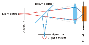 Principle of confocal microscopy.
In a confocal laser scanning microscope, a laser beam passes through a light source aperture and then is focused by an objective lens into a small (ideally diffraction limited) focal volume within or on the surface of a specimen. In biological applications especially, the specimen may be fluorescent. Scattered and reflected laser light as well as any fluorescent light from the illuminated spot is then re-collected by the objective lens. A beam splitter separates off some portion of the light into the detection apparatus, which in fluorescence confocal microscopy will also have a filter that selectively passes the fluorescent wavelengths while blocking the original excitation wavelength. After passing a pinhole, the light intensity is detected by a photodetection device (usually a photomultiplier tube (PMT) or avalanche photodiode), transforming the light signal into an electrical one that is recorded by a computer.[4]
The detector aperture obstructs the light that is not coming from the focal point, as shown by the dotted gray line in the image. The out-of-focus light is suppressed: most of the returning light is blocked by the pinhole, which results in sharper images than those from conventional fluorescence microscopy techniques and permits one to obtain images of planes at various depths within the sample (sets of such images are also known as z stacks).[1]
The detected light originating from an illuminated volume element within the specimen represents one pixel in the resulting image. As the laser scans over the plane of interest, a whole image is obtained pixel-by-pixel and line-by-line, whereas the brightness of a resulting image pixel corresponds to the relative intensity of detected light. The beam is scanned across the sample in the horizontal plane by using one or more (servo controlled) oscillating mirrors. This scanning method usually has a low reaction latency and the scan speed can be varied. Slower scans provide a better signal-to-noise ratio, resulting in better contrast and higher resolution. Information can be collected from different focal planes by raising or lowering the microscope stage or objective lens. The computer can generate a three-dimensional picture of a specimen by assembling a stack of these two-dimensional images from successive focal planes.[1]
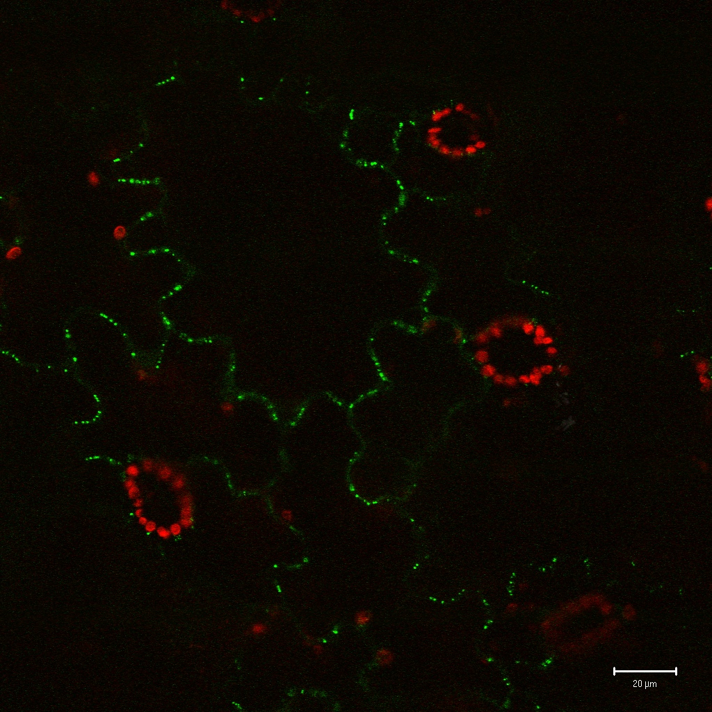
 An example of a GFP fusion protein.
Confocal microscopy provides the capacity for direct, noninvasive, serial optical sectioning of intact, thick, living specimens with a minimum of sample preparation as well as a marginal improvement in lateral resolution.[4] Biological samples are often treated with fluorescent dyes to make selected objects visible. However, the actual dye concentration can be low to minimize the disturbance of biological systems: some instruments can track single fluorescent molecules. Also, transgenic techniques can create organisms that produce their own fluorescent chimeric molecules (such as a fusion of GFP, green fluorescent protein with the protein of interest).
Resolution enhancement
CLSM is a scanning imaging technique in which the resolution obtained is best explained by comparing it with another scanning technique like that of the scanning electron microscope (SEM). CLSM has the advantage of not requiring a probe to be suspended nanometers from the surface, as in an AFM or STM, for example, where the image is obtained by scanning with a fine tip over a surface. The distance from the objective lens to the surface (called the working distance) is typically comparable to that of a conventional optical microscope. It varies with the system optical design, but working distances from hundreds of micrometres to several millimeters are typical.
In CLSM a specimen is illuminated by a point laser source, and each volume element is associated with a discrete scattering or fluorescence intensity. Here, the size of the scanning volume is determined by the spot size (close to diffraction limit) of the optical system because the image of the scanning laser is not an infinitely small point but a three-dimensional diffraction pattern. The size of this diffraction pattern and the focal volume it defines is controlled by the numerical aperture of the system's objective lens and the wavelength of the laser used. This can be seen as the classical resolution limit of conventional optical microscopes using wide-field illumination. However, with confocal microscopy it is even possible to improve on the resolution limit of wide-field illumination techniques because the confocal aperture can be closed down to eliminate higher orders of the diffraction pattern. For example, if the pinhole diameter is set to 1 Airy unit then only the first order of the diffraction pattern makes it through the aperture to the detector while the higher orders are blocked, thus improving resolution at the cost of a slight decrease in brightness. In fluorescence observations, the resolution limit of confocal microscopy is often limited by the signal to noise ratio caused by the small number of photons typically available in fluorescence microscopy. One can compensate for this effect by using more sensitive photodetectors or by increasing the intensity of the illuminating laser point source. Increasing the intensity of illumination laser risks excessive bleaching or other damage to the specimen of interest, especially for experiments in which comparison of fluorescence brightness is required. When imaging tissues which are differentially refractive, such as the spongy mesophyll of plant leaves or other air-space containing tissues, spherical aberrations that impair confocal image quality are often pronounced. Such aberrations however, can be significantly reduced by mounting samples in optically transparent, non-toxic perfluorocarbons such as perfluorodecalin, which readily infiltrates tissues and has a refractive index almost identical to that of water [1].
Uses
CLSM is widely-used in numerous biological science disciplines, from cell biology and genetics to microbiology and developmental biology.
Clinically, CLSM is used in the evaluation of various eye diseases, and is particularly useful for imaging, qualitative analysis, and quantification of endothelial cells of the cornea.[5] It is used for localizing and identifying the presence of filamentary fungal elements in the corneal stroma in cases of keratomycosis, enabling rapid diagnosis and thereby early institution of definitive therapy. Research into CLSM techniques for endoscopic procedures is also showing promise.[6] In the pharmaceutical industry, it was recommended to follow the manufacturing process of thin film pharmaceutical forms, to control the quality and uniformity of the drug distribution.[7] CLSM is also used as the data retrieval mechanism in some 3D optical data storage systems and has helped determine the age of the Magdalen papyrus.
See also
- Confocal microscopy
- LSM_(Zeiss) Laser Scanning Confocal Microscopy platform format form Carl Zeiss
- Two-photon excitation microscopy : Although they use a related technology (both are laser scanning microscopes), multiphoton fluorescence microscopes are not strictly confocal microscopes. The term confocal arises from the presence of a diaphragm in the conjugated focal plane (confocal). This diaphragm is usually absent in multiphoton microscopes.
- Total internal reflection fluorescence microscope (TIRF)
- Fluorescence microscope
- STED microscopy
- Point Spread Function
References
- ^ a b c d Pawley JB (editor) (2006). Handbook of Biological Confocal Microscopy (3rd ed.). Berlin: Springer. ISBN 038725921X.
- ^ US 3013467
- ^ Considerations on a laser-scanning-microscope with high resolution and depth of field: C. Cremer and T. Cremer in M1CROSCOPICA ACTA VOL. 81 NUMBER 1 September,pp. 31—44 (1978)
- ^ a b Fellers TJ, Davidson MW (2007). "Introduction to Confocal Microscopy". Olympus Fluoview Resource Center. National High Magnetic Field Laboratory. http://www.olympusconfocal.com/theory/confocalintro.html. Retrieved 2007-07-25.
- ^ Patel DV, McGhee CN (2007). "Contemporary in vivo confocal microscopy of the living human cornea using white light and laser scanning techniques: a major review". Clin. Experiment. Ophthalmol. 35 (1): 71–88. doi:10.1111/j.1442-9071.2007.01423.x. PMID 17300580.
- ^ Hoffman A, Goetz M, Vieth M, Galle PR, Neurath MF, Kiesslich R (2006). "Confocal laser endomicroscopy: technical status and current indications". Endoscopy 38 (12): 1275–83. doi:10.1055/s-2006-944813. PMID 17163333.
- ^ Le Person S, Puigalli JR, Baron M, Roques M Near infrared drying of pharmaceutical thin film: experimental analysis of internal mass transport Chemical Engineering and Processing 37 , 257-263 , 1998
External links
Categories:- Microscopes
- Cell imaging
- Scientific techniques

Wikimedia Foundation. 2010.