- Submandibular triangle
-
Submandibular triangle 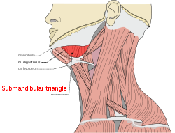
Submandibular triangle 
Side of neck, showing chief surface markings. (Nerves are yellow, arteries are red.) Latin trigonum submandibulare Gray's subject #145 564 The submandibular triangle (or submaxillary or digastric triangle) corresponds to the region of the neck immediately beneath the body of the mandible.
Contents
Boundaries and coverings
It is bounded:
- above, by the lower border of the body of the mandible, and a line drawn from its angle to the mastoid process;
- below, by the posterior belly of the Digastricus; in front, by the anterior belly of the Digastricus.
It is covered by the integument, superficial fascia, Platysma, and deep fascia, ramifying in which are branches of the facial nerve and ascending filaments of the cutaneous cervical nerve.
Its floor is formed by the Mylohyoideus.
Divisions
It is divided into an anterior and a posterior part by the stylomandibular ligament.
Anterior part
The anterior part contains the submandibular gland, superficial to which is the anterior facial vein, while imbedded in the gland is the external maxillary artery and its glandular branches.
Beneath the gland, on the surface of the Mylohyoideus, are the submental artery and the mylohyoid artery and nerve.
Posterior part
The posterior part of this triangle contains the external carotid artery, ascending deeply in the substance of the parotid gland
This vessel lies here in front of, and superficial to, the internal carotid, being crossed by the facial nerve, and gives off in its course the posterior auricular, superficial temporal, and internal maxillary branches: more deeply are the internal carotid, the internal jugular vein, and the vagus nerve, separated from the external carotid by the Styloglossus and Stylopharyngeus, and the hypoglossal nerve
See also
Additional images
-

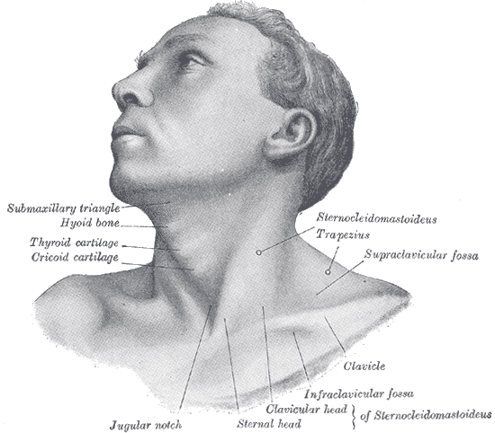
Anterolateral view of head and neck.
-
The triangles of the neck. (Anterior triangles to the left; posterior triangles to the right. Suprahyoid labeled at left.)
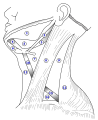
Summary of contents
There summarizes the important structures found in the submandibular triangle:
- 1. The external and internal carotid artery
- 2. The internal jugular vein
- 3. The deep cervical lymph nodes
- 4. The 10 th cranial nerve ( Vagus Nerve )
- 5. The submandibular gland
- 6. The submandibular lymph nodes
- 7. The Facial artery and vein
- 8. The 12 th cranial nerve ( Hypoglossal Nerve )
External links
- lesson5 at The Anatomy Lesson by Wesley Norman (Georgetown University) (necktriangle)
- lesson6 at The Anatomy Lesson by Wesley Norman (Georgetown University)
- SUNY Labs 25:16-0100
- SUNY Figs 25:01-01
- submandibular+triangle at eMedicine Dictionary
- Overview at bcm.edu
- Overview at howard.edu
This article was originally based on an entry from a public domain edition of Gray's Anatomy. As such, some of the information contained within it may be outdated.
Head Neck Triangles: Anterior of the neck (Muscular, Carotid, Submandibular, Submental) · Posterior of the neck (Occipital, Subclavian) · Suboccipital
Supraclavicular fossaThorax Triangle of auscultation · axillary folds (Anterior, Posterior) · Clavipectoral triangle · Inframammary fold
Infraclavicular fossaAbdomen/pelvis regions (Epigastrium, Hypochondrium, Umbilical region, Latus, Hypogastrium, Inguinal region)
quadrants (RUQ, LUQ, RLQ, LLQ)
McBurney's point · Desjardins' point · Traube's spacePerineal General anatomy: systems and organs, regional anatomy, planes and lines, superficial axial anatomy, superficial anatomy of limbsCategories:- Human anatomy
- Head and neck


Wikimedia Foundation. 2010.