- Carotid triangle
Infobox Anatomy
Name = PAGENAME
Latin = trigonum caroticum
GraySubject = 145
GrayPage = 564
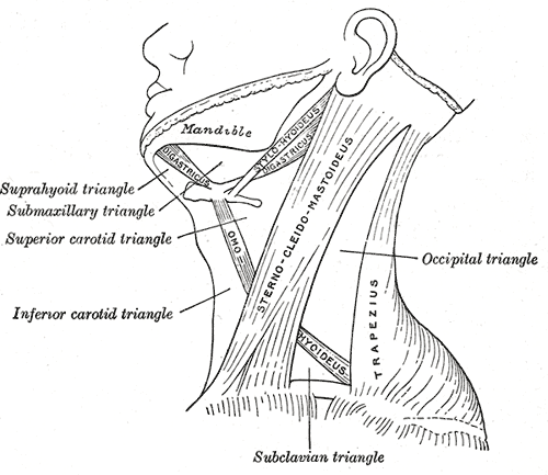
Caption = The triangles of the neck. (Anterior triangles to the left; posterior triangles to the right. Superior carotid triangle labeled at center left.)

Caption2 = Side of neck, showing chief surface markings. (Nerves are yellow, arteries are red.)
System =
MeshName =
MeshNumber =
DorlandsPre = t_19
DorlandsSuf = 12823361
The carotid triangle (or superior carotid triangle) is a portion of theanterior triangle of the neck .Coverings and boundaries
It is bounded:
* behind by theSternocleidomastoideus ;
* below, by the superior belly of theOmohyoideus
* above, by theStylohyoideus and the posterior belly of theDigastricus .It is covered by the integument, superficial fascia,
Platysma and deep fascia; ramifying in which are branches of the facial and cutaneous cervical nerves.Its floor is formed by parts of the
Thyrohyoideus ,Hyoglossus , and theConstrictores pharyngis medius and inferior.Arteries
This space when dissected is seen to contain the upper part of the common carotid artery, which bifurcates opposite the upper border of the
thyroid cartilage into the external and internal carotid. These vessels are somewhat concealed from view by the anterior margin of the Sternocleidomastoideus, which overlaps them.The external and internal carotids lie side by side, the external being the more anterior of the two.
The following branches of the external carotid are also met with in this space:
* thesuperior thyroid artery , running forward and downward;
* thelingual artery , directly forward;
* thefacial artery , forward and upward;
* theoccipital artery , backward;
* theascending pharyngeal artery , directly upward on the medial side of the internal carotid.Veins
The veins met with are:
* theinternal jugular , which lies on the lateral side of the common and internal carotid arteries;
* and veins corresponding to the above-mentioned branches of the external carotid—viz.,
** thesuperior thyroid ,
** thelingual ,
**common facial ,
**ascending pharyngeal ,
** and sometimes theoccipital ...all of which end in the internal jugular.
Nerves
The nerves in this space are the following.
In front of the sheath of the
common carotid is theramus descendens hypoglossi .The
hypoglossal nerve crosses both the internal and external carotids above, curving around the origin of theoccipital artery .Within the sheath, between the artery and vein, and behind both, is the
vagus nerve ; behind the sheath, thesympathetic trunk .On the lateral side of the vessels, the
accessory nerve runs for a short distance before it pierces theSternocleidomastoideus ; and on the medial side of theexternal carotid , just below thehyoid bone , may be seen the internal branch of thesuperior laryngeal nerve ; and, still more inferiorly, the external branch of the same nerve.Other contents
The upper portion of the larynx and lower portion of the pharynx are also found in the front part of this space.
ee also
*
Anterior triangle of the neck
=AdditionalExternal links
* (NormanAnatomyFig|necktriangle)
*
*
Wikimedia Foundation. 2010.