- Corniculate cartilages
-
Corniculate cartilages 
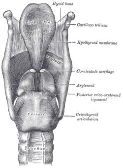
Ligaments of the larynx. Posterior view. (Corniculate cartilage labeled at center right.) 
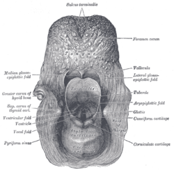
The entrance to the larynx, viewed from behind. (Corniculate cartilage labeled at bottom right.) Latin cartilagines corniculata Gray's subject #236 1075 The corniculate cartilages (cartilages of Santorini) are two small conical nodules consisting of yellow elastic cartilage, which articulate with the summits of the arytenoid cartilages and serve to prolong them posteriorly and medially.
They are situated in the posterior parts of the aryepiglottic folds of mucous membrane, and are sometimes fused with the arytenoid cartilages.
Contents
Eponym
It is named for Giovanni Domenico Santorini.[1][2]
Additional images
References
External links
This article was originally based on an entry from a public domain edition of Gray's Anatomy. As such, some of the information contained within it may be outdated.
Head and neck anatomy, Upper RT: Larynx (TA A06.2, TH H3.05.01, GA 11.1072) Cartilages major/unpaired: Epiglottis (Vallecula) · Thyroid (Laryngeal prominence, Oblique line, Superior thyroid notch, Superior horn, Inferior horn) · Cricoid
minor/paired: Arytenoid (Vocal process, Muscular process) · Corniculate · CuneiformLigaments/folds extrinsic ligaments: Hyoepiglottic ligament · Thyrohyoid membrane (Lateral ligament, Median ligament) · Thyroepiglottic ligament · Cricotracheal ligament
intrinsic ligaments · upper: Quadrangular membrane (Aryepiglottic, Vestibular ligament/Vestibular fold)
intrinsic ligaments · lower: Cricothyroid ligament (Median, Lateral/Conus elasticus, Vocal ligament/Vocal folds)Laryngeal cavity Other Categories:- Head and neck
- Respiratory system stubs
Wikimedia Foundation. 2010.


