- Cuneiform cartilages
-
Cuneiform cartilages 
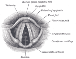
Laryngoscopic view of interior of larynx. 
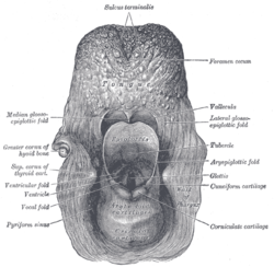
The entrance to the larynx, viewed from behind. Latin cartilagines cuneiformes Gray's subject #235 1075 The cuneiform cartilages of the larynx (cartilages of Wrisberg) are two small, elongated pieces of yellow elastic cartilage, placed one on either side, in the aryepiglottic fold, where they give rise to small whitish elevations on the surface of the mucous membrane, just in front of the arytenoid cartilages.
Additional images
This article was originally based on an entry from a public domain edition of Gray's Anatomy. As such, some of the information contained within it may be outdated.
Head and neck anatomy, Upper RT: Larynx (TA A06.2, TH H3.05.01, GA 11.1072) Cartilages major/unpaired: Epiglottis (Vallecula) · Thyroid (Laryngeal prominence, Oblique line, Superior thyroid notch, Superior horn, Inferior horn) · Cricoid
minor/paired: Arytenoid (Vocal process, Muscular process) · Corniculate · CuneiformLigaments/folds extrinsic ligaments: Hyoepiglottic ligament · Thyrohyoid membrane (Lateral ligament, Median ligament) · Thyroepiglottic ligament · Cricotracheal ligament
intrinsic ligaments · upper: Quadrangular membrane (Aryepiglottic, Vestibular ligament/Vestibular fold)
intrinsic ligaments · lower: Cricothyroid ligament (Median, Lateral/Conus elasticus, Vocal ligament/Vocal folds)Laryngeal cavity Other Categories:- Head and neck
- Respiratory system stubs
Wikimedia Foundation. 2010.


