Hepatorenal recess of subhepatic space
- Hepatorenal recess of subhepatic space
-
| Hepatorenal recess of subhepatic space |
 |
| Latin |
recessus hepatorenalis recessi subhepatici |
The hepatorenal recess[1] or subhepatic recess is the space that separates the liver from the right kidney. As a potential space, the recess is not filled with fluid under normal conditions. However, fluid can collect in this space in circumstances where the abdomen fills with fluid, such as hemoperitoneum. This fluid may be seen on ultrasound or computed tomography (CT scan).
Clinical importance
Since it is a potential space, the hepatorenal recess is not normally filled with fluid. However, this space becomes significant in conditions in which fluid collects within the abdomen (most commonly ascites and hemoperitoneum). The intraperitoneal fluid, be it blood, ascites, or dialysate, collects in this space and may be visualized, most commonly via ultrasound or computed tomography (CT) scanning. As little as 30 or 40 ml of fluid in the abdominal cavity may be visualized in this space.
Early visualization of fluid in the hepatorenal recess on FAST scan may be an indication for urgent laparotomy.[2]
Name
The hepatorenal recess is also called the pouch of Morison (or Morison's pouch) after the British surgeon James Rutherford Morison.[3]
References
- ^ Forbis, Pat; Bartolucci, Susan L.; Stedman, Thomas Lathrop (2005). Stedman's medical eponyms. Hagerstown, MD: Lippincott Williams & Wilkins. pp. p498. ISBN 0-7817-5443-7.
- ^ Weill F, Le Mouel A, Bihr E, Rohmer P, Zeltner F, Sauget Y (April 1980). "[Ultrasonic diagnosis of intraperitoneal fluid in Morison's pouch (and in the splenoperitoneal recess): the moon crescent sign (author's transl)]" (in French). J Radiol 61 (4): 251–6. PMID 7392002.
- ^ Morison's pouch at Who Named It?
Additional images
-
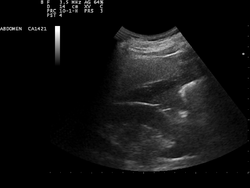
A normal ultrasonographic view of Morison's pouch. The bright line is the capsule of the kidney; there is no fluid present and hence no visible space.
-
Ultrasound of liver (left side of the image) and right kidney (right side of the image) and Morison's pouch, not containing fluid.
-
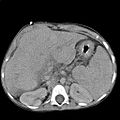
Abdominal CT, showing Morison's pouch as the dark margin surrounding the right kidney (at lower left corner of image).
-
Ultrasonographic view of the abdomen demonstrating fluid within Morison's pouch.
-
Morison's pouch with fluid present (at red arrows).
-
Wikimedia Foundation.
2010.
Look at other dictionaries:
Hepatorenal recess — The hepatorenal recesscite book |author=Forbis, Pat; Bartolucci, Susan L.; Stedman, Thomas Lathrop |title=Stedman s medical eponyms |publisher=Lippincott Williams Wilkins |location=Hagerstown, MD |year=2005 |pages=p498 |isbn=0 7817 5443 7 |oclc=… … Wikipedia
recess — A small hollow or indentation. SYN: recessus [TA]. [L. recessus] anterior r. a circumscript deepening of the interpeduncular fossa in the direction of the mamillary bodies. SYN: recessus anterior [TA]. anterior r. of tympanic … Medical dictionary
pouch — A pocket or cul de sac. SEE ALSO: fossa, recess, sac. antral p. a p. made in the antrum of the stomach of experimental animals. branchial pouches SYN: pharyngeal pouches. Broca p. SYN: puden … Medical dictionary
recessus — SYN: recess. [L. a withdrawing, a receding] r. anterior [TA] SYN: anterior recess. r. anterior membranae tympanicae [TA] SYN: anterior recess of tympanic membrane. r. cochlearis [TA] SYN: cochlear recess. r. costodiaphragmaticus [TA] SYN:… … Medical dictionary
Recto-uterine pouch — Sagittal section of the lower part of a female trunk, right segment. (Excavatio recto uterina labeled at bottom right.) … Wikipedia
Abdomen — Female Belly In vertebrates such as mammals the abdomen (belly) constitutes the part of the body between the thorax (chest) and pelvis. The region enclosed by the abdomen is termed the abdominal cavity. In arthropods it is the most distal section … Wikipedia
Ovarian ligament — Ligament: Ovarian ligament Uterus and right broad ligament, seen from behind. The broad ligament has been spread out and the ovary drawn downward. The ligament of ovary is labeled at the center top. The suspensory ligament of the ovary (not… … Wikipedia
True ribs — Anterior surface of sternum and costa cartilages. (True ribs are first seven.) Gray s subject #28 123 The first seven ribs ar … Wikipedia
Mesometrium — Uterus and right broad ligament, seen from behind. (Broad ligament visible at center. Mesometrium constitutes majority of the broad ligament.) The mesometrium is the mesentery of the uterus. It constitutes the majority of the broad ligament of… … Wikipedia
Cystohepatic triangle — The cystic artery branches from the right hepatic artery. Re … Wikipedia


