- High-content screening
-
High Content screening is an automated cell biology method drawing on optics, chemistry, biology and image analysis to permit rapid, highly parallel biological research and drug discovery.
Contents
General principles
High-content screening is a drug discovery method that uses images of living cells as the basic unit for molecule discovery. Proteins of interest present in the cells are detected using fluorescent tags, such as the green fluorescent protein, or by fluorescent antibodies. Image analysis is then used to measure changes in properties of the cells caused by external treatment such as chemical inhibitors or RNA interference- for example if the cells division is slowed or entry of a protein into the cell is arrested. High content screening is the combination of modern cell biology, with all its molecular tools, with automated high resolution microscopy and robotic handling. It differs from most life science work because experiment evaluation is mostly automated. The technology is mainly used to determine whether a potential drug affects aspects of cell function involved in or that describe a disease. For example, in humans G-protein coupled receptors (GPCRs) are a large family of around 880 cell surface proteins that transduce extra-cellular changes in the environment into a cell response, like triggering an increase in blood pressure because of the release of a regulatory hormone into the blood stream. Activation of these GPCRs can involve their entry into cells and when this can be visualised it can be the basis of a systematic analysis of receptor function through chemical genetics, systematic genome wide screening or physiological manipulation.
High Content screening is also known as cell based screening, phenotypic screening or visual screening, and it has a major place in pharmaceutical company drug discovery. All these terms refer to the systematic search for new drugs, small molecule inhibitors or chemical entities that could have use in biology or medicine. The parallel acquisition of data on different cell properties, for example activity of signal transduction cascades and cytoskeleton integrity is the main advantage of this method in comparison to the faster but less detailed high throughput screening. While HCS is slower, the wealth of acquired data allows a more profound understanding of drug effects.
Automated image based screening permits the identification of small compounds altering cellular phenotypes and is of interest for the discovery of new pharmaceuticals and new cell biological tools for modifying cell function. The selection of molecules based on a cellular phenotype does not require a priori knowledge of the biochemical targets that are affected by compounds and while this may be a benefit for compound discovery, the biochemical target itself must be subsequently identified. Given the increase in the use of phenotypic/visual screening as a cell biological tool, methods are required that permit systematic biochemical target identification if these molecules are to be of broad use (Burdine and Kodadek, 2004). Target identification has been defined as the rate limiting step in chemical genetics/high-content screening (Eggert and Mitchison, 2006).
 The goal of HCS is better research and new pharmaceutical discovery
The history of high-content screening
High-content screening technology allows for the evaluation of multiple biochemical and morphological parameters in cellular systems. Through combining the imaging of cells in microtiter plates with automated image analysis algorithms, researchers can acquire deeper knowledge on multiple biochemical or morphological pathways at the single-cell level at an early stage in the development new drugs.
The utility of automated cell biology requires an examination of how automation and objective measurement can improve the experimentation and the understanding of disease. First, it removes the influence of the investigator in most, but not all, aspects of cell biology research and second it makes entirely new approaches to possible.
In review, classical 20th century cell biology used cell lines grown in culture where the experiments were measured using very similar to that described here, but there the investigator made the choice on what was measured and how. In the early 1990s, the development of CCD cameras (charge coupled device cameras) for research created the opportunity to measure features in pictures of cells- such as how much protein is in the nucleus, how much is outside. Sophisticated measurements soon followed using new fluorescent molecules, which are used to measure cell properties like second messenger concentrations or the pH of internal cell compartments. The wide use of the green fluorescent protein, a natural fluorescent protein molecule from jellyfish, then accelerated the trend toward cell imaging as a mainstream technology in cell biology. Despite these advances, the choice of which cell to image and which data to present and how to analyse it was still selected by the investigator.
By analogy, if one imagines a football field and dinner plates laid across it, instead of looking at all of them, the investigator would choose a handful near the score line and had to leave the rest. In this analogy the field is a tissue culture dish, the plates the cells growing on it. While this was a reasonable and pragmatic approach automation of the whole process and the analysis makes possible the analysis of the whole population of living cells, so the whole football field can be measured.
From one to many—what this offers for research
This technology allows a (very) large number of experiments to be performed, allowing explorative screening. Currently its main use is in chemical genetics where large, diverse small molecule collections are systematically tested for their effect on cellular model systems. Novel drugs can be found using screens of tens of thousands of molecules, and these have promise for the future of drug development. Beyond drug discovery, chemical genetics is aimed at functionalizing the genome by identifying small molecules that acts on most of the 21,000 gene products in a cell. High-content technology will be part of this effort which could provide useful tools for learning where and when proteins act by knocking them out chemically. This would be most useful for gene where knock out mice (missing one or several genes) can not be made because the protein is required for development, growth or otherwise lethal when it is not there. Chemical knock out could address how and where these genes work. Further the technology is used in combination with RNAi to identify sets of genes involved in specific mechanisms, for example cell division. Here, libraries of RNAis, covering a whole set of predicted genes inside the target organisms genome can be used to identify relevant subsets, facilitating the annotation of genes for which no clear role has been established beforehand. The large datasets produced by automated cell biology contain spatially resolved, quantitative data which can be used for building for systems level models and simulations of how cells and organisms function. Systems biology models of cell function would permit prediction of why, where and how the cell responds to external changes, growth and disease.
High-content screening technology
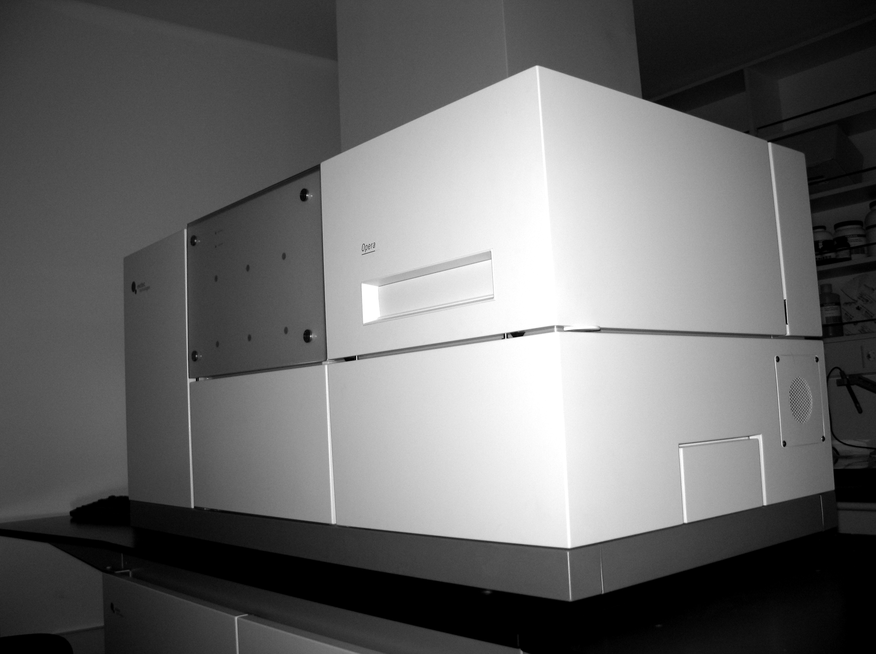
 An automated confocal image reader
High-content screening technology is mainly based on automated digital microscopy and flow cytometry, in combination with IT-systems for the analysis and storage of the data. “High-content” or visual biology technology has two purposes, first to acquire spatially or temporally resolved information on an event and second to automatically quantify it. Spatially resolved instruments are typically automated microscopes, and temporal resolution still requires some form of fluorescence measurement in most cases.This means that a lot of HCS instruments are (fluorescence) microscopes that are connected to some form of image analysis package. These take care of all the steps in taking fluorescent images of cells and provide rapid, automated and unbiased assessment of experiments.
Technology providers
The instruments on the market can be divided on the basis of price, footprint and the ethereal design qualities of the box they come in - but the most incisive difference is whether the instruments are optical confocal or not. Confocal imaging summarizes as imaging/resolving a thin slice through an object and rejecting out of focus light that comes from outside this slide. This gives higher image signal to noise and higher resolution than the more commonly applied epi-fluorescence microscopy. For many biological assays, confocal imaging is not ideal (e.g. phototoxicity issues or the need for a larger focal depth etc.). What all instruments share is the ability to take, store and interpret images automatically and most integrate into large robotic cell/medium handling platforms.
- The current confocal platforms are the point scanning 4 color ImageXpress ULTRA (Molecular Devices, Union City, USA), the spinning disk (nipkow disk) Pathway 855 and 435 from BD Biosciences (formerly Atto Biosciences, Rockville, Maryland), Opera (PerkinElmer Inc., Waltham, MA) and the slit scanning IN Cell 3000 (GE/Amersham Biosciences, Cardiff, UK).
- The current widefield platforms are: Arrayscan VTI (Cellomics (Cellomics)), IN Cell Analyzer 2000 (GE Healthcare Piscataway, New Jersey, USA), Acumen eX3 (TTP LabTech Ltd, Cambridge, UK), Scanalyzer (Scanalyzer LemnaTec, Aachen Germany) and "ImageXpress MICRO" (Molecular Devices, Union City, USA). Several instruments are ready for live cell temperature controlled imaging - such as the "IN Cell 1000" (GE/Amersham Biosciences Piscataway, New Jersey, USA), the "Pathway HT" (Becton Dickinson Biosciences) and the "ImageXpress MICRO" (Molecular Devices, Union City, USA), "Scan^R" (Olympus Soft Imaging Solutions, Germany), which may be of use to biologists interested in following events over relatively long time courses in vivo.
Alternatively more generic microscope instrumentation such as the Cell Observer by Carl Zeiss can be used for high content screening. While such instruments are less specialized they can be more appealing to academic settings, where tasks and experiments change more rapidly than in industrial research.
Additionally, many of these HCS platforms have been extended by (Caliper life sciences (Caliper Life Science))'s (Hopkinton, MA) integration of liquid handlers Sciclone G3 Liquid Handling Workstation and Zephyr, as well as plate moving robot Twister II to further automate the screening process.
Dedicated software for image analysis is available from these vendors (IN Cell Investigator, Perkin Elmer Columbus) or from specialised firms such as DCILabs (Belgium), LemnaTec (Germany) and from Accerlys Pipeline Pilot, as well as from some groups which provide open-source software for image analysis, such as BioImageXD, CellProfiler, and ImageJ. Dedicated open-source software for data management and data mining: Open source web based database system to publish image based screening experiments HCPB.
Kits for high-content screening of various target proteins (e.g. p53, c-jun and NFkB) have recently become available from commercial suppliers.
Timeline of the evolution of the science and technology
- (5/96) Introduction of the BDI High Content Screening System ArrayScan system introduces high throughput cell-based screening
- (10/96) Founding of Cellomics
- (1999) Introduction of the Cellomics ArrayScan II
- (1999) Introduction of the Q3DM EIDAQ 100 High Throughput Microscope
- (1999) Introduction of the Universal Imaging Gen-1
- (1999) Introduction of the Acumen Explorer
- (2000) Introduction of the Universal Imaging Discovery-1
- (date) Introduction of the IN Cell Analyzer 3000
- (date) Introduction of the Evotec/PerkinElmer Opera [1]
- (date) Introduction of the PerkinElmer Operetta [2]
- (2010) Introduction of the PerkinElmer Columbus
- (date) Introduction of the IN Cell Analyzer 1000
- (date) Introduction of Transfluor assay
- (date) Introduction of the Atto Pathway HT
- (date) Introduction of Axon Instruments ImageXpress 5000
- (1999) Introduction of Scanalyzer Platform Scanalyzer
- (5/05) Introduction of the Molecular Devices ImageXpress Micro [3]
- (01/06) Introduction of the TTP LabTech Acumen eX3
- (1/06) Introduction of the Molecular Devices ImageXpress Ultra [4]
- (3/06) Introduction of the Olympus scan^R
- 48h life cell imaging on cell arrays - Life cell imaging with 1536 experiments in parallel.
- Cell detecting autofocus system with memory function
- (01/08) Introduction of the Acclerys Pipeline Pilot Platform for HCS Informatics
- (03/09) Introduction of the IN Cell Analyzer 2000
- (12/10) Introduction of the Vala Sciences IC200 Kinetic Image Cytometer
- (9/11) Introduction of the Molecular Devices ImageXpress Micro XL Widefield HCS System
See also
References
- Abraham VC, Taylor DL, Haskins JR., High-content screening applied to large-scale cell biology, Trends Biotechnol. 2004 Jan;22(1):15-22. PMID 14690618.
- Bleicher KH, Bohm HJ, Muller K, Alanine AI., Hit and lead generation: beyond high-throughput screening, Nat Rev Drug Discov. 2003 May;2(5):369-78. PMID 12750740.
- Burdine, L., and T. Kodadek. 2004. Target identification in chemical genetics: the (often) missing link. Chem Biol. 11:593-7. PMID 15157870.
- Carpenter AE, Sabatini DM, Systematic genome-wide screens of gene function, Nature Reviews Genetics, 2004, 5(1):11-22. PMID 14708012.
- Edwards BS, Oprea T, Prossnitz ER, Sklar LA., Flow cytometry for high-throughput, high-content screening, Curr Opin Chem Biol. 2004 Aug;8(4):392-8. PMID 15288249.
- Eggert, U.S., and T.J. Mitchison. 2006. Small molecule screening by imaging. Curr Opin Chem Biol. PMID 16682248.
- Mawji IA, Xu GW, Macrae CJ, Koch CA, Datti A, Wrana JL, Dennis JW, Schimmer AD.A high-content chemical screen identifies ellipticine as a modulator of p53 nuclear localization. Apoptosis. 2008 Mar;13(3):413-22. PMID 18181020.
- Giuliano KA, Haskins JR, Taylor DL, Advances in high-content screening for drug discovery, Assay Drug Dev Technol. 2003 Aug;1(4):565-77. PMID 15090253.
- Milligan G., High-content assays for ligand regulation of G-protein-coupled receptors., Drug Discov Today. 2003 Jul 1;8(13):579-85. PMID 12850333.
External links
- RNAiAtlas RNAi Libraries and their target analysis results (ETH Zurich)
- The Advanced Cell Classifier project (ETH Zurich)
- LabAutopedia
- High Content Imaging Google Group (High Content Imaging Users Group)
- Yale Center for High Throughput Cell Biology (Yale Center for High Throughput Cell Biology)
- High-Content Screening (Purdue University)
- High-Throughput Bioscience Center (HTBC, Stanford University)
- Institut Pasteur Korea
- BioHts HCS page
- Max Planck Institute of Molecular Cell Biology and Genetics (MPI-CBG)
- Molecular Screening Shared Resource (MSRI,UCLA)
- Central Drug Research Institute, Lucknow, India
- Open Screening Environment
- HCS browser and HCS-based classification
- Screening Centre KIT Karlsruhe Institute of Technology / Forschungszentrum Karlsruhe
- [5] Burnham Institute for Medical Research / High Content Screening Core Facility
- [6] Cambridge Healthtech Institute
- Image Mining Group @ Institut Pasteur Korea
Categories:

Wikimedia Foundation. 2010.