- Minimally differentiated acute myeloblastic leukemia
-
Minimally differentiated acute myeloblastic leukemia Classification and external resources 
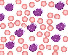
A schematic showing the appearance of acute myeloblastic leukemia, M0 under microscope. Blasts show no Auer rods.ICD-O: M9872/3 MeSH D015470 Minimally differentiated acute myeloblastic leukemia is a subtype of AML. It is classified as M0 by FAB. It represents 2–3% of all cases of AML.[1] Although minimally differentiated AML was recognized earlier, criteria for FAB M0 were developed in 1991.[2] The blasts in these cases cannot be recognized as myeloid based on morphology and cytochemistry, but immunophenotyping demonstrates myeloid antigens.[3]
Cytochemistry and Molecular Features
In acute myeloblastic leukemia (M0), the blasts are agranular and nonreactive when stained for myeloperoxidase activity, and Auer rods are not seen. The blasts react with antibodies to myeloperoxidase and antibodies to CD13, CD33, and CD34. Human leukocyte antigen (HLA)-DR is positive in most patients. Occasional cases require in situ hybridization to identify the myeloperoxidase gene315 or genomic profiling for early myeloid-associated genes.[4] Abnormal and unfavorable karyotypes (e.g., loss of the long arm of chromosome 5 (5q-) and 7q-) and higher expression of the multidrug resistance glycoprotein (p170) are frequent.[5] In general, minimally differentiated acute myeloblastic leukemia has a poor prognosis.[6]
References
- ^ Kumar V, Abbas AK, Fausto N, Mitchel RN (2007). Robbins basic Pathology (8th ed.). Saunders.
- ^ Lee EJ, Pollak A, Leavitt RD, Testa JR, Schiffer CA (November 1987). "Minimally differentiated acute nonlymphocytic leukemia: a distinct entity". Blood 70 (5): 1400–6. PMID 3663939. http://www.bloodjournal.org/cgi/pmidlookup?view=long&pmid=3663939.
- ^ Greer JP, Foerster J, Lukens JN (2003). Wintrobe's Clinical Hematology (11th ed.). Lippincott Williams & Wilkins.
- ^ Testa U, Torelli GF, Riccioni R, et al. (June 2002). "Human acute stem cell leukemia with multilineage differentiation potential via cascade activation of growth factor receptors". Blood 99 (12): 4634–7. doi:10.1182/blood.V99.12.4634. PMID 12036900. http://www.bloodjournal.org/cgi/pmidlookup?view=long&pmid=12036900.
- ^ Lichtman M., et al. (2005). Williams Hematology (7th ed.). McGraw-Hill.
- ^ Cuneo A, Ferrant A, Michaux JL, et al. (June 1995). "Cytogenetic profile of minimally differentiated (FAB M0) acute myeloid leukemia: correlation with clinicobiologic findings". Blood 85 (12): 3688–94. PMID 7780152. http://www.bloodjournal.org/cgi/pmidlookup?view=long&pmid=7780152.
See also
- Acute myeloid leukemia
- Acute myeloblastic leukemia without maturation (M1 in FAB) — M9873/3
- Acute myeloblastic leukemia with maturation (M2 in FAB) — M9874/3
Myeloid hematological malignancy/leukemia histology (ICD-O 9590–9989, C81–C96, 200–208) CFU-GM/
and other granulocytesCFU-GMOtherCFU-BasoCFU-EosMEP CFU-MegCFU-EMD (Refractory anemia, Refractory anemia with excess of blasts, Chromosome 5q deletion syndrome, Sideroblastic anemia, Paroxysmal nocturnal hemoglobinuria, Refractory cytopenia with multilineage dysplasia)CFU-Mast Mastocytosis: Diffuse cutaneous mastocytosis · Erythrodermic mastocytosis · Generalized eruption of cutaneous mastocytosis (adult type) · Generalized eruption of cutaneous mastocytosis (childhood type) · Mast cell sarcoma · Solitary mastocytoma · Systemic mastocytosis · Xanthelasmoidal mastocytosisMultiple/unknown Categories:
Wikimedia Foundation. 2010.
