- Nile blue
-
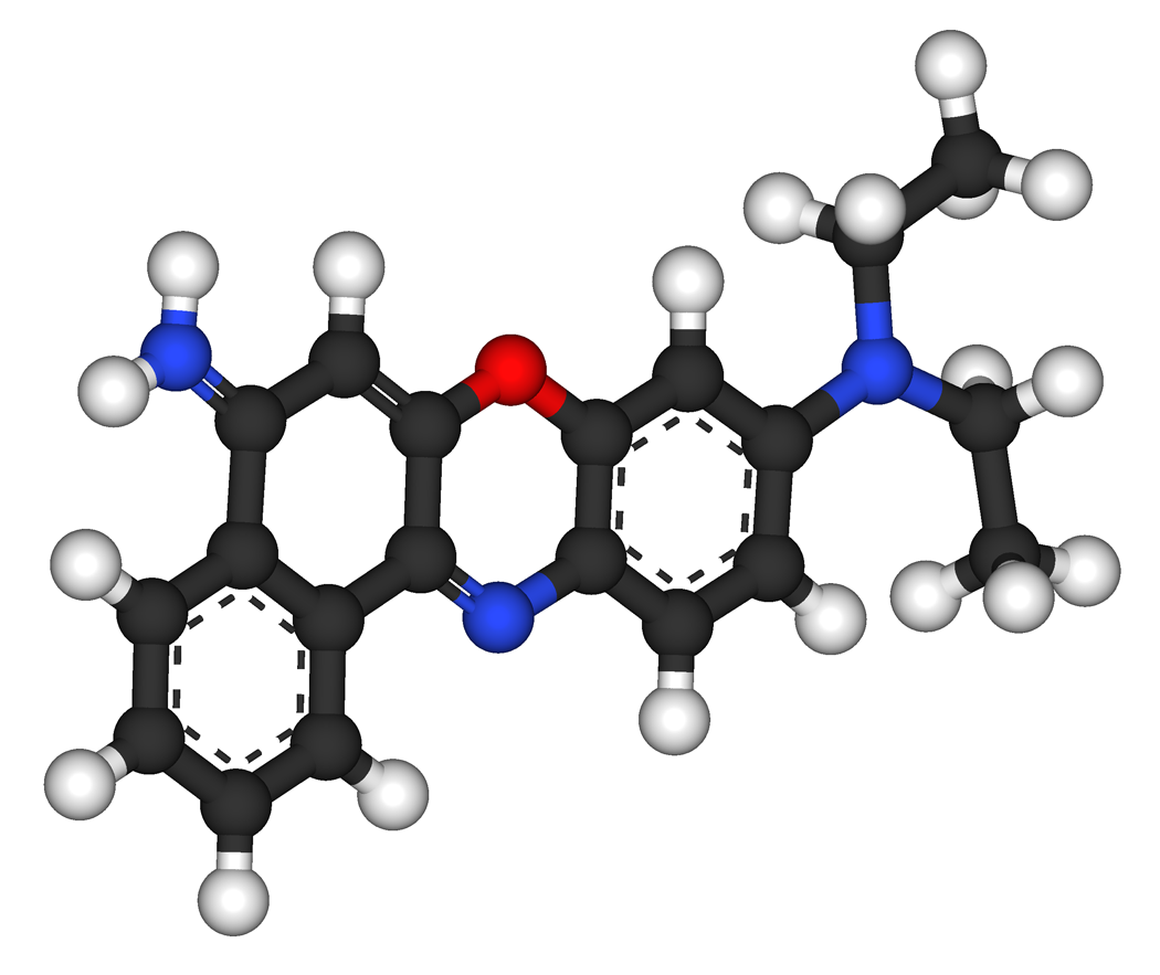
Nile blue 
 Nile blueOther namesNile blue A
Identifiers CAS number 3625-57-8 PubChem 422690 ChemSpider 374117 
Jmol-3D images Image 1 - N=1c3c(OC=2C=1\C=C/C(N(CC)CC)C=2)cc(c4c3cccc4)N
- InChI=1S/C20H21N3O/c1-3-23(4-2)13-9-10-17-18(11-13)24-19-12-16(21)14-7-5-6-8-15(14)20(19)22-17/h5-13H,3-4,21H2,1-2H3
Key: WOIORDFNOALMFO-UHFFFAOYSA-N
InChI=1/C20H21N3O/c1-3-23(4-2)13-9-10-17-18(11-13)24-19-12-16(21)14-7-5-6-8-15(14)20(19)22-17/h5-13H,3-4,21H2,1-2H3
Key: WOIORDFNOALMFO-UHFFFAOYAF
Properties Molecular formula C20H20ClN3O Molar mass 353.845 g/mol  blue (verify) (what is:
  /
/ ?)
?)
Except where noted otherwise, data are given for materials in their standard state (at 25 °C, 100 kPa)Infobox references
Nile blue (or Nile blue A) is a stain used in biology and histology. It may be used with live or fixed cells, and imparts a blue colour to cell nuclei.It may also be used in conjunction with fluorescence microscopy to stain for the presence of polyhydroxybutyrate granules in prokaryotic or eukaryotic cells. Boiling a solution of Nile blue with sulfuric acid produces Nile red (Nile blue oxazone).
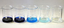 Nile blue hydrochloride in water.
Concentrations, left to right: 1000 ppm, 100 ppm, 10 ppm, 1 ppm, 100 ppb. Nile blue in water.
Left to right: pH 0, pH 4, pH 7, pH 10, pH 14.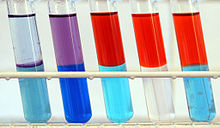 Nile blue in water (lower phase) and ethyl acetate (upper phase) in daylight.
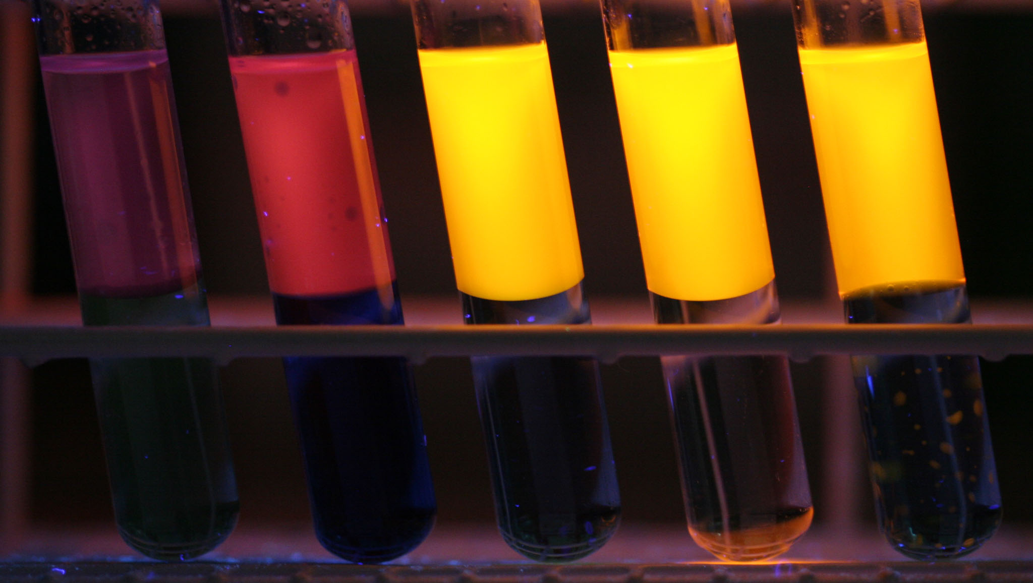
Left to right: pH 0, pH 4, pH 7, pH 10, pH 14 Nile blue in water (lower phase) and ethyl acetate (upper phase) in UV light (366 nm).
Left to right: pH 0, pH 4, pH 7, pH 10, pH 14 Nile blue (free base) in daylight (top row) and UV light (366 nm, bottom row) in different solvents.
Left to right: 1. methanol, 2. ethanol, 3. methyl-tert-butylether, 4. cyclohexane, 5. n-hexane, 6. acetone, 7. tetrahydrofuran, 8. ethyl acetate, 9. dimethyl formamide, 10. acetonitrile, 11. toluene, 12. chloroformContents
Chemical and physical properties
Nile blue is a fluorescent dye. The fluorescence shows especially in nonpolar solvents with a high quantum yield.[1]
The absorption and emission maxima of Nile blue are strongly dependent on pH and the solvents used:[1]
Solvent Absorption λ max (nm) Emission λ max (nm) Toluene 493 574 Acetone 499 596 Dimethylformamide 504 598 Chloroform 624 647 1-Butanol 627 664 2-propanol 627 665 Ethanol 628 667 Methanol 626 668 Water 635 674 1.0 N hydrochloric acid (pH = 1.0) 457 556 0.1 N sodium hydroxide solution (pH = 11.0) 522 668 Ammonia water (pH = 13.0) 524 668 The duration of Nile blue fluorescence in ethanol was measured as 1.42 ns. This is shorter than the corresponding value of Nile red with 3.65 ns. The fluorescence duration is independent on dilution in the range 10−3–10−8 mol/L.[1]
Nile blue staining
Nile blue is used for histological staining of biological preparations. It highlights the distinction between neutral lipids (triglycerides, cholesteryl esters, steroids) which are stained pink and acids (fatty acids, chromolipids, phospholipids) which are stained blue.[2]
The Nile blue staining, according to Kleeberg, uses the following chemicals:
- Nile Blue A
- 1% acetic acid
- Glycerol or glycerol gelatin
Workflow
The sample or frozen sections is/are fixated in formaldehyde, then immersed for 20 minutes in the Nile blue solution and rinsed with water. For better differentiation, it is dipped in 1% acetic acid for 10–20 minutes until the colors are pure. This might take only 1–2 minutes. Then the sample is thoroughly rinsed in water (for one to two hours). Afterwards, the stained specimen is taken on a microscope slide and excess water is removed. The sample can be embedded in glycerol or glycerol gelatin.
Results
Unsaturated glycerides are pink, nuclei and elastins(?) dark, fatty acids and various fatty substances and fat mixtures are purple blue.[3]
Example: Detection of poly-β-hydroxybutyrate granules (PHB)
The PHB granules in the cells of Pseudomonas solanacearum can be visualized by Nile blue A staining. The PHB granules in the stained smears are observed with an epifluorescence microscope under oil immersion, at a 1000 times magnification; under 450 nm excitation wavelength they show a strong orange fluorescence.[4]
Nile blue in DNA Electrophoresis
Nile blue is also apparently used in a variety of commercial DNA staining formulations used for DNA electrophoresis.[5] As it does not require UV trans-illumination in order to be visualised in an agarose gel as with ethidium bromide, it can be used to observe DNA as it is separated and also as a dye to aid in gel-extraction of DNA fragments without incurring damage by UV-irradiation.
Nile blue in oncology
Derivatives of Nile blue are potential photosensitizers in photodynamic therapy of malignant tumors. These dyes aggregate in the tumor cells, especially in the lipid membranes, and/or are sequestered and concentrated in subcellular organelles.[6]
With the Nile blue derivative N-Ethyl-Nile Blue (EtNBA), normal and premalignant tissues in animal experiments can be distinguished by fluorescence spectroscopy in fluorescence imaging. EtNBA shows no phototoxic effects.[7]
References
- ^ a b c Jose, Jiney; Burgess, Kevin (2006). "Benzophenoxazine-based fluorescent dyes for labeling biomolecules". Tetrahedron 62 (48): 11021. doi:10.1016/j.tet.2006.08.056. http://www.chem.tamu.edu/cfi/Publications/221.pdf.
- ^ Roche Lexikon, accessed 25 June 2007.
- ^ Benno Romeis, Mikroskopische Technik, 15. Aufl., R. Oldenbourg Verlag, München, 1948
- ^ 97/647/EG: Entscheidung der EU-Kommission vom 9. September 1997 über ein vorläufiges Versuchsprogramm für Diagnose, Nachweis und Identifizierung von Pseudomonas solanacearum (Smith) Smith in Kartoffeln, accessed 27 June 2007.
- ^ PDF DNA staining protocol for schools, University of Reading
- ^ Lin, CW; Shulok, JR; Kirley, SD; Cincotta, L; Foley, JW (1991). "Lysosomal localization and mechanism of uptake of Nile blue photosensitizers in tumor cells". Cancer research 51 (10): 2710–9. PMID 2021950.
- ^ Van Staveren, HJ; Speelman, OC; Witjes, MJ; Cincotta, L; Star, WM (2001). "Fluorescence imaging and spectroscopy of ethyl nile blue a in animal models of (pre)malignancies". Photochemistry and photobiology 73 (1): 32–8. doi:10.1562/0031-8655(2001)073<0032:FIASOE>2.0.CO;2. PMID 11202363.
External links
Categories:- Fluorescent dyes
- Oxazin dyes
- Vital stains
- Naphthobenzoxazines


Wikimedia Foundation. 2010.