- Pi helix
-
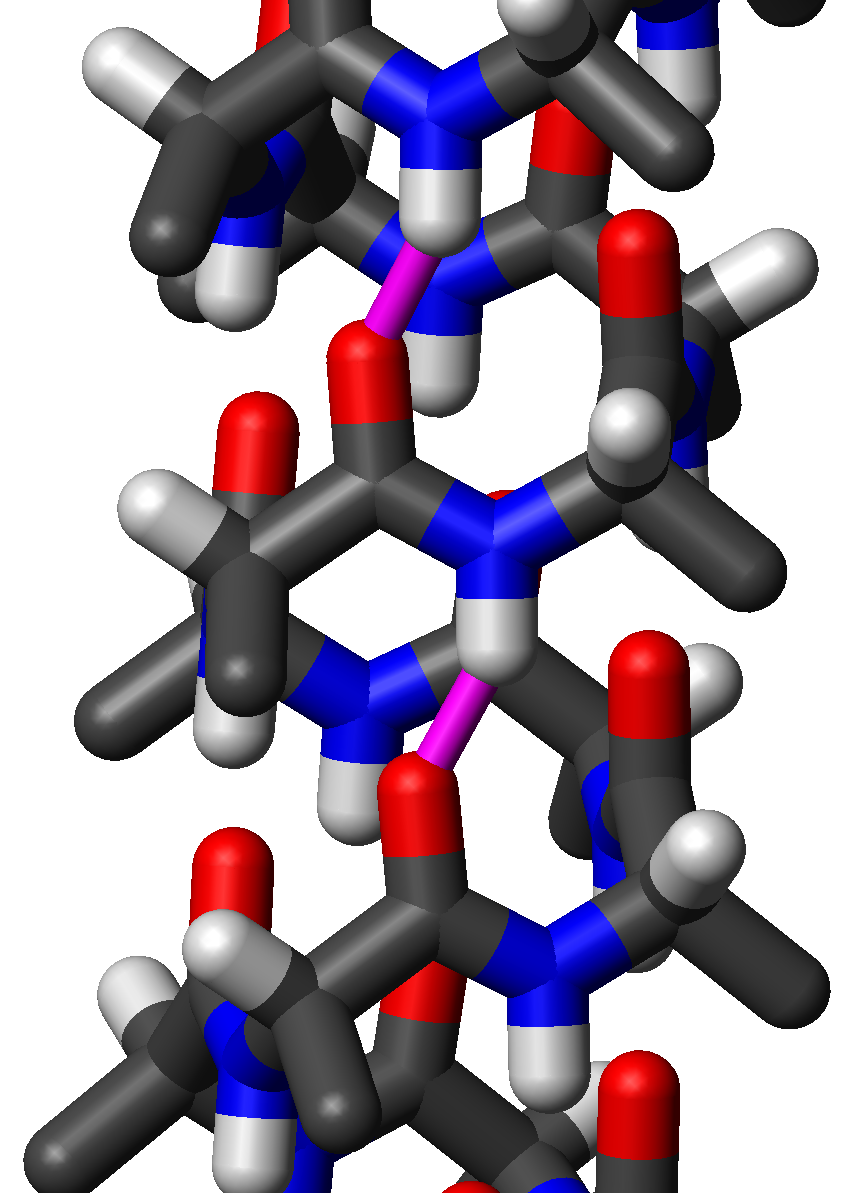
 Side view of a standard π-helix of L-alanine residues in atomic detail. Two hydrogen bonds to the same peptide group are highlighted in magenta; the oxygen-hydrogen distance is 1.65 Å (165 pm). The protein chain runs upwards, i.e., its N-terminus is at the bottom and its C-terminus at the top of the figure. Note that the sidechains point slightly downwards, i.e., towards the N-terminus.
A pi helix (or π-helix) is a type of secondary structure found in proteins.[1] Although thought to be rare, π-helices are actually found in 15% of known protein structures and are believed to be an evolutionary adaptation derived by the insertion of a single amino acid into an α-helix.[2] Because such insertions are highly destabilizing,[3] the formation of π-helices would tend to be selected against unless it provided some functional advantage to the protein. π-helices therefore are typically found near functional sites of proteins. [2] [4] [5]
Contents
Standard structure
The amino acids in a standard π-helix are arranged in a right-handed helical structure. Each amino acid corresponds to a 87° turn in the helix (i.e., the helix has 4.1 residues per turn), and a translation of 1.15 Å (=0.115 nm) along the helical axis. Most importantly, the N-H group of an amino acid forms a hydrogen bond with the C=O group of the amino acid five residues earlier; this repeated i+5→i hydrogen bonding defines a π-helix. Similar structures include the 310 helix (i+3→i hydrogen bonding) and the α-helix (i+4→i hydrogen bonding).
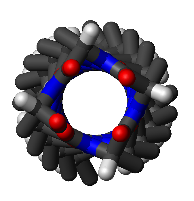
 Top view of the same helix shown above. Four carbonyl groups are pointing upwards towards the viewer, spaced roughly 87° apart on the circle, corresponding to 4.1 amino-acid residues per turn of the helix.
The majority of π-helices are only 7 residues in length and do not adopt regularly repeating (φ, ψ) dihedral angles throughout the entire structure like that of α-helices or ß-sheets. Because of this, textbooks that provide single dihedral values for all residues in the π-helix are misleading. Some generalizations can be made, however. When the first and last residue pairs are excluded, dihedral angles exist such that the ψ dihedral angle of one residue and the φ dihedral angle of the next residue sum to roughly -125°. The first and last residue pairs sum to -95° and -105°, respectively. For comparison, the sum of the dihedral angles for a 310 helix is roughly -75°, whereas that for the α-helix is roughly -105°. Proline is often seen immediately following the end of π-helices. The general formula for the rotation angle Ω per residue of any polypeptide helix with trans isomers is given by the equation
Left-handed structure
In principle, a left-handed version of the π-helix is possible by reversing the sign of the (φ, ψ) dihedral angles to (55°, 70°). This pseudo-"mirror-image" helix has roughly the same number of residues per turn (4.1) and helical pitch (1.5 angstroms or 150 picometers). It is not a true mirror image, because the amino-acid residues still have a left-handed chirality. A long left-handed π-helix is unlikely to be observed in proteins because, among the naturally occurring amino acids, only glycine is likely to adopt positive φ dihedral angles such as 55°.
See also
- alpha helix
- 3_10 helix
- secondary structure
References
- ^ Pauling L, Corey RB, Branson HR (1951). "The structure of proteins; two hydrogen-bonded helical configurations of the polypeptide chain". Proc. Nat. Acad. Sci. Wash. 37 (4): 205–211. doi:10.1073/pnas.37.4.205. PMC 1063337. PMID 14816373. http://www.pubmedcentral.nih.gov/articlerender.fcgi?tool=pmcentrez&artid=1063337.
- ^ a b Cooley RB, Arp DJ, Karplus PA (2010). "Evolutionary Origin of a Secondary Structure: pi-Helices as Cryptic but Widespread Insertional Variations of alpha-Helices That Enhance Protein Functionality.". J Mol Biol 404 (2): 232–246. doi:10.1016/j.jmb.2010.09.034. PMC 2981643. PMID 20888342. http://www.pubmedcentral.nih.gov/articlerender.fcgi?tool=pmcentrez&artid=2981643.
- ^ Keefe LJ, Sondek J, Shortle D, and Lattman EE (2000). "The alpha aneurism: a structural motif revealed in an insertion mutant of staphylococcal nuclease.". Proc Natl Acad Sci USA 90 (8): 3275–3279. PMC 46282. PMID 8475069. http://www.pubmedcentral.nih.gov/articlerender.fcgi?tool=pmcentrez&artid=46282.
- ^ Weaver TM (2000). "The pi-helix translates structure into function.". Protein science 9 (1): 201–206. PMC 2144447. PMID 10739264. http://www.pubmedcentral.nih.gov/articlerender.fcgi?tool=pmcentrez&artid=2144447.
- ^ Fodje MN, Al-Karadaghi S (2002). "Occurrence, conformational features and amino acid propensities for the pi-helix.". Protein Eng 15 (5): 353–358. PMID 12034854.
Protein secondary structure Protein secondary structure Helices: α-helix · 310 helix · π-helix · β-helix · Polyproline helix · Collagen helix
Extended: β-strand · Turn · Beta hairpin · Beta bulge · α-strand
Supersecondary: Coiled coil · Helix-turn-helix · EF handAmino acids Helix-favoring: Methionine · Alanine · Leucine · Glutamic acid · Glutamine · Lysine
Extended-favoring: Threonine · Isoleucine · Valine · Phenylalanine · Tyrosine · Tryptophan
Disorder-favoring: Glycine · Serine · Proline · Asparagine · Aspartic acid
No preference: Cysteine · Histidine · Arginine←Primary structureTertiary structure→
This protein-related article is a stub. You can help Wikipedia by expanding it.
![3 \cos \Omega = 1 - 4 \cos^{2} \left[ \left(\phi + \psi \right)/2 \right]](5/8f504de02eb71fe4fabe5d44c83da475.png)