- Two-streams hypothesis
-
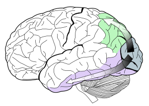
The two-streams hypothesis is a widely accepted, but still controversial, account of visual processing. As visual information exits the occipital lobe, it follows two main channels, or "streams". The ventral stream (also known as the "what pathway") travels to the temporal lobe and is involved with object identification. The dorsal stream (or, "where pathway") terminates in the parietal lobe and process spatial locations.
The hypothesis was originally proposed by Leslie Ungerleider and Mortimer Mishkin in 1982[1] and reviewed by George Ettlinger in 1990[2]. An influential early review on the subject was published by Melvyn Goodale and David Milner in 1992.[3]
However, recent experimental work has undermined the empirical basis of the dissociation,[4][5] and a recent review has concluded that it is "difficult if not impossible to test" and that, contrary to the two-streams hypothesis, a more "general and parsimonious one posits the existence of one single processing stream."[6]
Contents
Dorsal stream
The dorsal stream is proposed to be involved in the guidance of actions and recognizing where objects are in space. Also known as the parietal stream, the "where" stream, or the "how" stream, this pathway stretches from the primary visual cortex (V1) in the occipital lobe forward into the parietal lobe. It is interconnected with the parallel ventral stream (the "what" stream) which runs downward from V1 into the temporal lobe.
General features
The dorsal stream is involved in spatial awareness and guidance of actions (e.g., reaching). In this it has two distinct functional characteristics—it contains a detailed map of the visual field, and is also good at detecting and analyzing movements.
The dorsal stream commences with purely visual functions in the occipital lobe before gradually transferring to spatial awareness at its termination in the parietal lobe.
The posterior parietal cortex is essential for "the perception and interpretation of spatial relationships, accurate body image, and the learning of tasks involving coordination of the body in space".[7]
It contains individually functioning lobules, one area of which contains neurons that produce enhanced activation when attention is moved onto the stimulus or the animal saccades towards to a visual stimulus (LIP), and another section where visual and somatosensory information are integrated (VIP).
Effects of damage or lesions
Damage to the posterior parietal cortex causes a number of spatial disorders including:
- Simultanagnosia: where the patient can only describe single objects without the ability to perceive it as a component of a set of details or objects in a context (as in a scenario, e.g. the forest for the trees).
- Optic ataxia: where the patient can't use visuospatial information to guide arm movements.
- Hemispatial neglect: where the patient is unaware of the contralesional half of space (that is, they are unaware of things in their left field of view and focus only on objects in the right field of view; or appear unaware of things in one field of view when they perceive them in the other). For example, a person with this disorder may draw a clock, and then label it from 12, 1, 2, ..., 6, but then stop and consider their drawing complete.
- Akinetopsia: inability to perceive motion.
- Apraxia: inability to produce discretionary or volitional movement in the absence of muscular disorders.
Ventral stream
The ventral stream is associated with object recognition and form representation. It has strong connections to the medial temporal lobe (which stores long-term memories), the limbic system (which controls emotions), and the dorsal stream (which deals with object locations and motion).
The ventral stream gets its main input from the parvocellular (as opposed to magnocellular) layer of the lateral geniculate nucleus of the thalamus. These neurons project to V1 sublayers 4Cβ, 4A, 3B and 2/3a[8] successively. From there, the ventral pathway goes through V2 and V4 to areas of the inferior temporal lobe: PIT (posterior inferotemporal), CIT (central inferotemporal), and AIT (anterior inferotemporal). Each visual area contains a full representation of visual space. That is, it contains neurons whose receptive fields together represent the entire visual field. Visual information enters the ventral stream through the primary visual cortex and travels through the rest of the areas in sequence.
Moving along the stream from V1 to AIT, receptive fields increase their size, latency, and the complexity of their tuning.
All the areas in the ventral stream are influenced by extraretinal factors in addition to the nature of the stimulus in their receptive field. These factors include attention, working memory, and stimulus salience. Thus the ventral stream does not merely provide a description of the elements in the visual world—it also plays a crucial role in judging the significance of these elements.
Problems with the hypothesis
The dichotomy of the dorsal/ventral pathways is still contentious among scientists. The neuroscience community is still not in agreement regarding the degree to which these streams are segregated (they are, in fact, heavily interconnected[9]) or the functional significance which should be attached to them. The hypothesis is probably an over-simplification of the true state of affairs in the visual cortex and a recent review concluded that the hypothesis is "difficult if not impossible to test."[6]
Evidence from illusions
Much support comes from the findings that visual illusions such as the Ebbinghaus illusion may distort judgements of a perceptual nature, but when the subject responds with an action, such as grasping, no distortion occurs. However, recent work[4][5] suggests that both the action and perception systems are equally fooled by such illusions.
Eye movements versus action
Much support for the dichotomy comes from research that compares eye movements to actions such as grasping or reaching. Differences between the two data sets are claimed to support the hypothesis, but eye movements themselves are actions and not necessarily indicative of what is occurring perceptually. The reasoning behind many studies could therefore be considered flawed.[6]
References
- ^ Mishkin M, Ungerleider LG. (1982). "Contribution of striate inputs to the visuospatial functions of parieto-preoccipital cortex in monkeys.". Behav Brain Res. 6 (1): 57–77. doi:10.1016/0166-4328(82)90081-X. PMID 7126325.
- ^ Ettlinger G. (1990). ""Object vision" and "spatial vision": the neuropsychological evidence for the distinction.". Cortex 26 (3): 319–41. PMID 2123426.
- ^ Goodale MA, Milner AD (1992). "Separate visual pathways for perception and action". Trends Neurosci. 15 (1): 20–5. doi:10.1016/0166-2236(92)90344-8. PMID 1374953.
- ^ a b Franz VH, Gegenfurtner KR, Bülthoff HH, Fahle M. (2000). "Grasping visual illusions: no evidence for a dissociation between perception and action.". Psychol Sci. 11 (1): 20–5. doi:10.1111/1467-9280.00209. PMID 11228838.
- ^ a b Franz VH, Scharnowski F, Gegenfurtner (2005). "Illusion effects on grasping are temporally constant not dynamic.". J Exp Psychol Hum Percept Perform. 31 (6): 1359–78. doi:10.1037/0096-1523.31.6.1359. PMID 16366795.
- ^ a b c Cardoso-Leite, Pedro and Gorea, Andrei (2010). "On the Perceptual/Motor Dissociation: A Review of Concepts, Theory, Experimental Paradigms and Data Interpretations". Seeing and Perceiving 23 (2): 89–151. doi:10.1163/187847510X503588. PMID 20550823. http://brill.publisher.ingentaconnect.com/content/brill/sp/2010/00000023/00000002/art00001.
- ^ Mark F Bear, Barry Connors, Michael Paradiso, (2007). Neuroscience: Exploring the Brain. Hagerstown, MD: Lippincott Williams & Wilkins. ISBN 0-7817-6003-8.
- ^ Lamme, Sup�r H, Spekreijse H (1998). "Feedforward, horizontal, and feedback processing in the visual cortex". Current Opinion in Neurobiology 8 (4): 529–535. doi:10.1016/S0959-4388(98)80042-1. PMID 9751656.
- ^ Farivar R. (2009). "Dorsal-ventral integration in object recognition.". Brain Res Rev. 61 (2): 144–53. doi:10.1016/j.brainresrev.2009.05.006. PMID 19481571.
Wikimedia Foundation. 2010.

