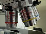
Multiphoton fluorescence microscope

Multiphoton fluorescence microscope

A multiphoton fluorescence microscope (MFM) is a specialized optical microscope.

Contents

Description

The MFM uses pulsed long-wavelength light to excite fluorophores within the specimen being observed. The fluorophore absorbs the energy from two long-wavelength photons which must arrive simultaneously in order to excite an electron into a higher energy state, from which it can decay, emitting a fluorescence signal. It differs from traditional fluorescence microscopy in which the excitation wavelength is shorter than the emission wavelength, as the summed energies of two long-wavelength exciting photons will produce an emission wavelength shorter than the excitation wavelength.

Multiphoton fluorescence microscopy has similarities to confocal laser scanning microscopy. Both use focused laser beams scanned in a raster pattern to generate images, and both have an optical sectioning effect. Unlike confocal microscopes, multiphoton microscopes do not contain pinhole apertures, which give confocal microscopes their optical sectioning quality. The optical sectioning produced by multiphoton microscopes is a result of the point spread function formed where the pulsed laser beams coincide. The multiphoton point spread function is typically dumbbell-shaped (longer in the x-y plane), compared to the upright rugby-ball shaped point spread function of confocal microscopes.

The longer wavelength, low energy (typically infra-red) excitation lasers of multiphoton microscopes are well-suited to use in imaging live cells as they cause less damage than short-wavelength lasers, so cells may be observed for longer periods with fewer toxic effects. Many researchers are currently working toward better and higher resolution multiphoton imaging developments.

See also

References

External links


Wikimedia Foundation. 2010.

Игры ⚽ Нужно сделать НИР?

Look at other dictionaries:

  • Fluorescence microscope — An upright fluorescence microscope (Olympus BX61) with the fluorescent filter cube turret above the objective lenses, coupled with a digital camera. A fluorescence microscope is an optical microscope used to study properties of organic or… …   Wikipedia

  • microscope — /muy kreuh skohp /, n. 1. an optical instrument having a magnifying lens or a combination of lenses for inspecting objects too small to be seen or too small to be seen distinctly and in detail by the unaided eye. 2. (cap.) Astron. the… …   Universalium

  • Multifocal multiphoton microscopy — is a microscopy technique for generating 3D images. By exploiting specific properties of pulsed mode multiphoton excitation the conflict between the density of the foci, i.e. the degree of parallelization, and the axial sectioning has been… …   Wikipedia

  • Near-field scanning optical microscope — Near field scanning optical microscopy (NSOM/SNOM) is a microscopic technique for nanostructure investigation that breaks the far field resolution limit by exploiting the properties of evanescent waves. This is done by placing the detector very… …   Wikipedia

  • Two-photon excitation microscopy — is a fluorescence imaging technique that allows imaging of living tissue up to a very high depth, that is up to about one millimeter. Being a special variant of the multiphoton fluorescence microscope, it uses red shifted excitation light which… …   Wikipedia

  • Optical sectioning — is the process by which a suitably designed microscope can produce clear images of a focal planes deep within a thick sample. This is used to reduce the need for thin sectioning using instruments such as the microtome. Many different techniques… …   Wikipedia

  • Microscopy — is the technical field of using microscopes to view samples and objects that cannot be seen with the unaided eye (objects that are not within the resolution range of the normal eye). There are three well known branches of microscopy, optical,… …   Wikipedia

  • Confocal laser scanning microscopy — (CLSM or LSCM) is a technique for obtaining high resolution optical images with depth selectivity.[1] The key feature of confocal microscopy is its ability to acquire in focus images from selected depths, a process known as optical sectioning.… …   Wikipedia

  • Multiphotonenmikroskopie — Zweiphotonen Fluoreszenzaufnahme an einem Schnitt durch einen Mausdarm. Zellkerne in grün, Schleim der Becherzellen in blau, Aktin (Phalloidin Färbung) in rot. Anregung erfolgte be …   Deutsch Wikipedia

  • Zwei-Photonen-Mikroskop — Zweiphotonen Fluoreszenzaufnahme an einem Schnitt durch einen Mausdarm. Zellkerne in grün, Schleim der Becherzellen in blau, Aktin (Phalloidin Färbung) in rot. Anregung erfolgte bei 780 nm durch einen Titan:Saphir Laser …   Deutsch Wikipedia

Share the article and excerpts

Direct link
Do a right-click on the link above
and select “Copy Link”