- Transversus thoracis muscle
-
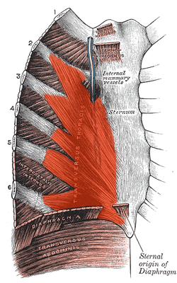
Transversus thoracis muscle Posterior surface of sternum and costal cartilages, showing Transversus thoracis. Latin musculus transversus thoracis Gray's subject #117 403 Origin costal cartilages of last 3-4 ribs, body of sternum, xiphoid process Insertion ribs/costal cartilages 2-6 Artery intercostal arteries/veins Nerve intercostal nerves Actions depresses ribs The transversus thoracis lies internal to the thoracic cage, anteriorly. It is a thin plane of muscular and tendinous fibers, situated upon the inner surface of the front wall of the chest. It is in the same layer as the subcostal muscles and the innermost intercostal muscles.
It arises on either side from the lower third of the posterior surface of the body of the sternum, from the posterior surface of the xiphoid process, and from the sternal ends of the costal cartilages of the lower three or four true ribs.
Its fibers diverge upward and lateralward, to be inserted by slips into the lower borders and inner surfaces of the costal cartilages of the second, third, fourth, fifth, and sixth ribs.
The lowest fibers of this muscle are horizontal in their direction, and are continuous with those of the Transversus abdominis; the intermediate fibers are oblique, while the highest are almost vertical.
This muscle varies in its attachments, not only in different subjects, but on opposite sides of the same subject.
The muscle is supplied by the anterior rami of the Thoracic spinal nerves (intercostal nerves).
Function
It is almost completely without function, but it separates the thoracic cage from the parietal pleura.
Contraction of this muscle aids in exertional expiration by decreasing the transverse diameter of the thoracic cage.
Additional images
External links
- LUC tth
- Transversus+thoracis+muscle at eMedicine Dictionary
- SUNY Labs 18:05-0104 - "Thoracic Wall: Removal of Intercostal Muscles"
- thoraxmuscles at The Anatomy Lesson by Wesley Norman (Georgetown University)
This article was originally based on an entry from a public domain edition of Gray's Anatomy. As such, some of the information contained within it may be outdated.
Back splenius (capitis, cervicis) · erector spinae (iliocostalis, longissimus, spinalis) · latissimus dorsi
transversospinales: (semispinalis dorsi, semispinalis cervicis, semispinalis capitis, multifidus, rotatores) · interspinales · intertransversarii
Vertebral column: trapezius · latissimus dorsi · rhomboid (major, minor) · levator scapulae
fascia: Thoracolumbar fasciaThorax intercostales (external, internal, innermost) · subcostales · transversus thoracis · levatores costarum · serratus posterior (inferior, superior) · diaphragm
Thoracic cavity: pectoralis major · pectoralis minor · subclavius · serratus anterior
fascia: Pectoral fascia · Clavipectoral fasciaCategories:- Muscle stubs
- Muscles of the torso
Wikimedia Foundation. 2010.


