- Immunoglobulin light chain
-
 Schematic diagram of an typical antibody showing two Ig heavy chains (blue) linked by disulfide bonds to two Ig light chains (green). The constant (C) and variable (V) domains are shown.
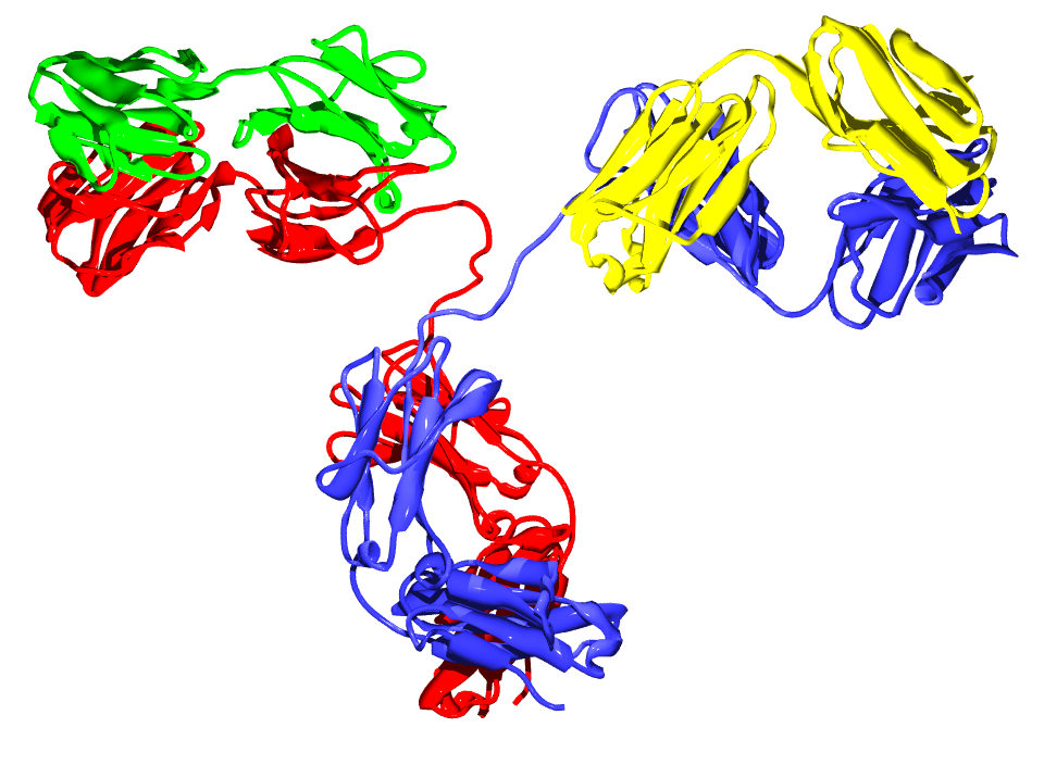
 An antibody molecule. The two heavy chains are colored red and blue and the two light chains green and yellow. See also:[1]
The immunoglobulin light chain is the small polypeptide subunit of an antibody (immunoglobulin).
A typical antibody is composed of two immunoglobulin (Ig) heavy chains and two Ig light chains.
Contents
In humans
There are two types of light chain in humans (as in other mammals),
- kappa (κ) chain, encoded by the immunoglobulin kappa locus (IGK@) on chromosome 2
- lambda (λ) chain, encoded by the immunoglobulin lambda locus (IGL@) on chromosome 22
Antibodies are produced by B lymphocytes, each expressing only one class of light chain. Once set, light chain class remains fixed for the life of the B lymphocyte. In a healthy individual, the total kappa to lambda ratio is roughly 2:1 in serum (measuring intact whole antibodies) or 1:1.5 if measuring free light chains, with a highly divergent ratio indicative of neoplasm.
The exact normal ratio of kappa to lambda ranges from 0.26 to 1.65. Both the kappa and the lambda chains can increase proportionately, maintaining a normal ratio. This is usually indicative of something other than a blood cell dyscrasia, such as kidney disease.
In other animals
The immunoglobulin light chain genes in tetrapods can be classified into three distinct groups: kappa (κ), lambda (λ), and sigma (σ). The divergence of the κ, λ, and σ isotypes preceded the radiation of tetrapods. The σ isotype was lost after the evolution of the amphibian lineage and before the emergence of the reptilian lineage.[1]
Other types of light chains can be found in lower vertebrates, such as the Ig-Light-Iota chain of Chondrichthyes and Teleostei.[2][3]
Camelids are unique among mammals as they have fully functional antibodies which have two heavy chains, but lack the light chains usually paired with each heavy chain.[4] The functional role of this separate repertoire is unknown as yet.
Structure
Only one type of light chain is present in a typical antibody, thus the two light chains of an individual antibody are identical.
Each light chain is composed of two tandem immunoglobulin domains:
- one constant (CL) domain
- one variable domain (VL) that is important for binding antigen
The approximate length of a light chain protein is from 211 to 217 amino acids.[2]
In pathology
Ig light chains produced in neoplastic plasma cells, e.g. in multiple myeloma, are called Bence Jones proteins.
See also
References
- ^ Das S, Nikolaidis N, Klein J, Nei M (2008). "Evolutionary redefinition of immunoglobulin light chain isotypes in tetrapods using molecular markers". Proc Natl Acad Sci U S A. 105 (43): 16647–52. doi:10.1073/pnas.0808800105. PMC 2575474. PMID 18940927. http://www.pubmedcentral.nih.gov/articlerender.fcgi?tool=pmcentrez&artid=2575474.
- ^ a b Janeway CA, Jr. et al. (2001). Immunobiology. (5th ed.). Garland Publishing. (electronic full text via NCBI Bookshelf) ISBN 0-8153-3642-X.
- ^ IMGT Index Antibodies (or Immunoglobulins).
- ^ Hamers-Casterman C, Atarhouch T, Muyldermans S, Robinson G, Hamers C, Songa E, Bendahman N, Hamers R (1993). "Naturally occurring antibodies devoid of light chains". Nature 363 (6428): 446–8. doi:10.1038/363446a0. PMID 8502296.
External links
Antibodies Antibodies see also disorders of globin and globulin proteins
B proteins: BY STRUCTURE: membrane, globular (en, ca, an), fibrous
This protein-related article is a stub. You can help Wikipedia by expanding it.

![[1]](http://www.emc.maricopa.edu/faculty/farabee/BIOBK/ANTIBODY.gif)