- Extensor retinaculum of the hand
-
Extensor retinaculum of the hand 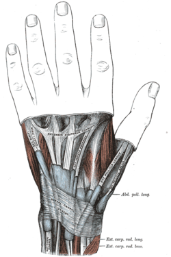
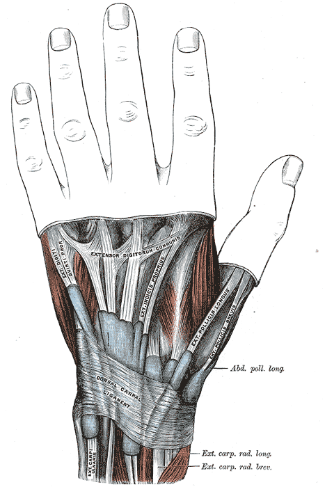
The mucous sheaths of the tendons on the back of the wrist. (Dorsal carpal ligament labeled at bottom center.) Latin retinaculum musculorum extensorum manus Gray's subject #126 458 The extensor retinaculum (dorsal carpal ligament, or posterior annular ligament) is an anatomical term for the thickened part of the antebrachial fascia that holds the tendons of the extensor muscles in place. It is located on the back of the forearm, just proximal to the hand. It is continuous with the palmar carpal ligament, which is located on the anterior side of the forearm.
It is a strong, fibrous band, extending obliquely downward and medialward across the back of the wrist, and consisting of part of the deep fascia of the back of the forearm, strengthened by the addition of some transverse fibers.
The extensor retinaculum is attached laterally to the lateral margin of the radius. However, it is not attached to the ulna medially, as the distance between these two bones varies with supination and pronation of the forearm. Instead the medial attachment is to the most medial of the carpal bones, the triquetrum (or triquetral bone) and pisiformis (or pisiform bone). The retinaculum is also attached in its passage across the wrist, to the ridges on the dorsal surface of the radius.
Additional images
-
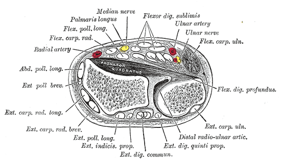
Transverse section across distal ends of radius and ulna.
External links
List of muscles of upper limbs (TA A04.6, GA 4.432) Shoulder deltoid · rotator cuff (supraspinatus, infraspinatus, teres minor, subscapularis) · teres major
fascia: Deltoid fascia · Supraspinous fascia · Infraspinous fasciaArm
(compartments)OtherForearm OtherHand Lateral volarMedial volarhypothenar (opponens digiti minimi, flexor digiti minimi brevis, abductor digiti minimi) · palmaris brevisIntermediateposterior: Extensor retinaculum · Extensor expansion
anterior: Flexor retinaculum · Palmar aponeurosisCategories:- Musculoskeletal system stubs
- Human anatomy
-
Wikimedia Foundation. 2010.