- dUTP diphosphatase
-
dUTP diphosphatase Identifiers EC number 3.6.1.23 CAS number 37289-34-2 Databases IntEnz IntEnz view BRENDA BRENDA entry ExPASy NiceZyme view KEGG KEGG entry MetaCyc metabolic pathway PRIAM profile PDB structures RCSB PDB PDBe PDBsum Gene Ontology AmiGO / EGO Search PMC articles PubMed articles dUTPase 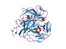
crystal structures of feline immunodeficiency virus dutp pyrophosphatase and its nucleotide complexes in three crystal forms. Identifiers Symbol dUTPase Pfam PF00692 Pfam clan CL0153 InterPro IPR008180 SCOP 1dup Available protein structures: Pfam structures PDB RCSB PDB; PDBe PDBsum structure summary dUTPase_2 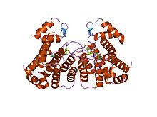
the crystal structure of a complex of campylobacter jejuni dutpase with substrate analogue dupnhp Identifiers Symbol dUTPase_2 Pfam PF08761 Pfam clan CL0231 InterPro IPR014871 SCOP 1w2y Available protein structures: Pfam structures PDB RCSB PDB; PDBe PDBsum structure summary In enzymology, a dUTP diphosphatase (EC 3.6.1.23) is an enzyme that catalyzes the chemical reaction
- dUTP + H2O
 dUMP + diphosphate
Thus, the two substrates of this enzyme are dUTP and H2O, whereas its two products are dUMP and diphosphate.
This enzyme belongs to the family of hydrolases, specifically those acting on acid anhydrides in phosphorus-containing anhydrides. The systematic name of this enzyme class is dUTP nucleotidohydrolase. Other names in common use include deoxyuridine-triphosphatase, dUTPase, dUTP pyrophosphatase, desoxyuridine 5'-triphosphate nucleotidohydrolase, and desoxyuridine 5'-triphosphatase. This enzyme participates in pyrimidine metabolism.
The main function of this enzyme is to remove dUTP from the deoxynucleotide pool, which reduces the probability of this base being mistakenly incorporated into DNA by DNA polymerases, which is a cause of mutations.[1]
Structural studies
As of late 2007, 48 structures have been solved for this class of enzymes, with PDB accession codes 1DUC, 1DUD, 1DUN, 1DUP, 1DUT, 1EU5, 1EUW, 1F7D, 1F7K, 1F7N, 1F7O, 1F7P, 1F7Q, 1F7R, 1MQ7, 1OGH, 1OGK, 1OGL, 1PKH, 1PKJ, 1PKK, 1RN8, 1RNJ, 1SEH, 1SIX, 1SJN, 1SLH, 1SM8, 1SMC, 1SNF, 1SYL, 1VYQ, 1W2Y, 2BSY, 2BT1, 2CJE, 2D4L, 2D4M, 2D4N, 2HQU, 2HR6, 2HRM, 2OKB, 2OKD, 2OKE, 2OL0, 2OL1, and 2PY4.
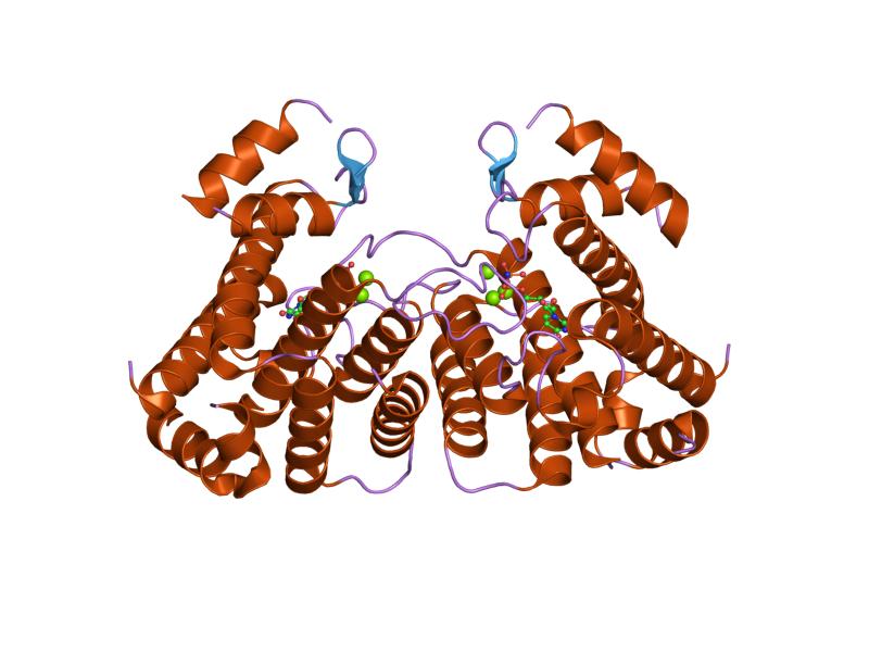
There are at least two structurally distinct families of dUTPases. The crystal structure of human dUTPase reveals that each subunit of the dUTPase trimer folds into an eight-stranded jelly-roll beta barrel, with the C-terminal beta strands interchanged among the subunits. The structure is similar to that of the Escherichia coli enzyme, despite low sequence homology between the two enzymes.[2]
The second family has a novel all-alpha fold, members of this family are unrelated to the all-beta fold found in dUTPases of the majority of organisms.[3]
References
- ^ Vassylyev DG, Morikawa K (1996). "Precluding uracil from DNA". Structure 4 (12): 1381–5. doi:10.1016/S0969-2126(96)00145-1. PMID 8994964.
- ^ Mol CD, Harris JM, McIntosh EM, Tainer JA (September 1996). "Human dUTP pyrophosphatase: uracil recognition by a beta hairpin and active sites formed by three separate subunits". Structure 4 (9): 1077–92. doi:10.1016/S0969-2126(96)00114-1. PMID 8805593.
- ^ Moroz, O. V.; Harkiolaki, M.; Galperin, M. Y.; Vagin, A. A.; González-Pacanowska, D.; Wilson, K. S. (2004). "The Crystal Structure of a Complex of Campylobacter jejuni dUTPase with Substrate Analogue Sheds Light on the Mechanism and Suggests the "Basic Module" for Dimeric d(C/U)TPases". Journal of Molecular Biology 342 (5): 1583–1597. doi:10.1016/j.jmb.2004.07.050. PMID 15364583.
Further reading
- Bertani Le., Haeggmark A., Reichard P. (1963). "Enzymatic Synthesis of Deoxyribonucleotides. II. Formation and Interconversion of Deoxyuridine Phosphates". J. Biol. Chem. 238: 3407–13. PMID 14085395.
- Giroir LE, Deutsch WA (1987). "Drosophila deoxyuridine triphosphatase. Purification and characterization". J. Biol. Chem. 262 (1): 130–4. PMID 3025197.
- Greenberg Gr., Somerville Rl. (1962). "DEOXYURIDYLATE KINASE ACTIVITY AND DEOXYURIDINETRIPHOSPHATASE IN ESCHERICHIA COLI". Proc. Natl. Acad. Sci. U. S. A. 48 (2): 247–57. doi:10.1073/pnas.48.2.247. PMC 220766. PMID 13901467. http://www.pubmedcentral.nih.gov/articlerender.fcgi?tool=pmcentrez&artid=220766.
- Grindey GR, Nichol CA (1971). "Mammalian deoxyuridine 5'-triphosphate pyrophosphatase". Biochim. Biophys. Acta. 240 (2): 180–3. PMID 5105331.
This article includes text from the public domain Pfam and InterPro IPR008180
This article includes text from the public domain Pfam and InterPro IPR014871

This hydrolase article is a stub. You can help Wikipedia by expanding it. - dUTP + H2O