- COQ7
-
The clk-1 (Clock abnormal protein 1) gene encodes an enzyme (demethoxyubiquinone mono-oxygenase) that is necessary for ubiquinone biosynthesis in the worm C. elegans and other eukaryotes. The mouse version of the gene is called mclk1 and the human, fruit fly and yeast homolog COQ7 (Coenzyme Q biosynthesis protein 7 homolog).[1][2]
Clk-1 is not to be confused with the unrelated CLK1 human gene which plays a role in RNA splicing.
Contents
Structure
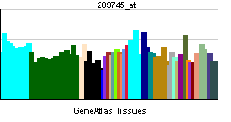
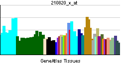
In 1999, Zoltan Vajo et al cloned COQ7 from human heart. They found that the human COQ7 protein contains 179 amino acids, is mostly helical, and contains an alpha-helical membrane insertion. It has a potential N-glycosylation site, a phosphorylation site for protein kinase C and another for casein kinase II, and 3 N-myristoylation sites. Northern blot analysis detected 3 transcripts; a 1-kb transcript was predominant in heart, and a 3-kb transcript was predominant in skeletal muscle, kidney, and pancreas.[3]
The protein has two repeats of about 90 amino acids, that contain two conserved motifs. One of these DXEXXH may be part of an enzyme active site. The structure and function of the gene are highly conserved among different species.[4]
The C. elegans protein contains 187 amino acid residues (20 kilodaltons), the human homolog 217 amino acid residues (24 kilodaltons, gene consisting of six exons spanning 11 kb and located on chromosome 16).[5]
Function
Ubiquinone is a small redox active lipid that is found in all membranes and that is a co-factor in numerous cellular redox processes, including mitochondrial electron transport. As a co-factor, ubiquinone is often involved in processes that produce reactive oxygen species (ROS). In addition, ubiquinone is one of the main endogenous antioxidants of the cell. The CLK-1 enzyme is responsible for the hydroxylation of 5-demethoxyubiquinone to 5-hydroxyubiquinone.
When ubiquinone biosynthesis is interrupted by the absence of the enzyme, cells accumulate an intermediate of ubiquinone biosynthesis, demethoxyubiquinone (DMQ).
It has been shown that mutations in the gene are associated with increased life span.[1][4] Defects of the gene slow down a variety of developmental and physiological processes, including the cell cycle, embryogenesis, post-embryonic growth, rhythmic behaviors and aging.[6]
References
- ^ a b Ewbank JJ, Barnes TM, Lakowski B, Lussier M, Bussey H, Hekimi S (February 1997). "Structural and functional conservation of the Caenorhabditis elegans timing gene clk-1". Science 275 (5302): 980–3. doi:10.1126/science.275.5302.980. PMID 9020081.
- ^ "Entrez Gene: COQ7 coenzyme Q7 homolog, ubiquinone (yeast)". http://www.ncbi.nlm.nih.gov/sites/entrez?Db=gene&Cmd=ShowDetailView&TermToSearch=10229.
- ^ Vajo Z, King LM, Jonassen T, Wilkin DJ, Ho N, Munnich A, Clarke CF, Francomano CA (October 1999). "Conservation of the Caenorhabditis elegans timing gene clk-1 from yeast to human: a gene required for ubiquinone biosynthesis with potential implications for aging". Mamm. Genome 10 (10): 1000–4. doi:10.1007/s003359901147. PMID 10501970.
- ^ a b Liu X, Jiang N, Hughes B, Bigras E, Shoubridge E, Hekimi S (October 2005). "Evolutionary conservation of the clk-1-dependent mechanism of longevity: loss of mclk1 increases cellular fitness and lifespan in mice". Genes Dev. 19 (20): 2424–34. doi:10.1101/gad.1352905. PMC 1257397. PMID 16195414. http://www.pubmedcentral.nih.gov/articlerender.fcgi?tool=pmcentrez&artid=1257397.
- ^ Asaumi S, Kuroyanagi H, Seki N, Shirasawa T (June 1999). "Orthologues of the Caenorhabditis elegans longevity gene clk-1 in mouse and human". Genomics 58 (3): 293–301. doi:10.1006/geno.1999.5838. PMID 10373327.
- ^ Felkai S, Ewbank JJ, Lemieux J, Labbé JC, Brown GG, Hekimi S (April 1999). "CLK-1 controls respiration, behavior and aging in the nematode Caenorhabditis elegans". EMBO J. 18 (7): 1783–92. doi:10.1093/emboj/18.7.1783. PMC 1171264. PMID 10202142. http://www.pubmedcentral.nih.gov/articlerender.fcgi?tool=pmcentrez&artid=1171264.
Further reading
- Maruyama K, Sugano S (1994). "Oligo-capping: a simple method to replace the cap structure of eukaryotic mRNAs with oligoribonucleotides.". Gene 138 (1-2): 171–4. doi:10.1016/0378-1119(94)90802-8. PMID 8125298.
- Suzuki Y, Yoshitomo-Nakagawa K, Maruyama K, et al. (1997). "Construction and characterization of a full length-enriched and a 5'-end-enriched cDNA library.". Gene 200 (1-2): 149–56. doi:10.1016/S0378-1119(97)00411-3. PMID 9373149.
- Jonassen T, Proft M, Randez-Gil F, et al. (1998). "Yeast Clk-1 homologue (Coq7/Cat5) is a mitochondrial protein in coenzyme Q synthesis.". J. Biol. Chem. 273 (6): 3351–7. doi:10.1074/jbc.273.6.3351. PMID 9452453.
- Asaumi S, Kuroyanagi H, Seki N, Shirasawa T (1999). "Orthologues of the Caenorhabditis elegans longevity gene clk-1 in mouse and human.". Genomics 58 (3): 293–301. doi:10.1006/geno.1999.5838. PMID 10373327.
- Vajo Z, King LM, Jonassen T, et al. (2000). "Conservation of the Caenorhabditis elegans timing gene clk-1 from yeast to human: a gene required for ubiquinone biosynthesis with potential implications for aging.". Mamm. Genome 10 (10): 1000–4. doi:10.1007/s003359901147. PMID 10501970.
- Hartley JL, Temple GF, Brasch MA (2001). "DNA cloning using in vitro site-specific recombination.". Genome Res. 10 (11): 1788–95. doi:10.1101/gr.143000. PMC 310948. PMID 11076863. http://www.pubmedcentral.nih.gov/articlerender.fcgi?tool=pmcentrez&artid=310948.
- Wiemann S, Weil B, Wellenreuther R, et al. (2001). "Toward a catalog of human genes and proteins: sequencing and analysis of 500 novel complete protein coding human cDNAs.". Genome Res. 11 (3): 422–35. doi:10.1101/gr.GR1547R. PMC 311072. PMID 11230166. http://www.pubmedcentral.nih.gov/articlerender.fcgi?tool=pmcentrez&artid=311072.
- Simpson JC, Wellenreuther R, Poustka A, et al. (2001). "Systematic subcellular localization of novel proteins identified by large-scale cDNA sequencing.". EMBO Rep. 1 (3): 287–92. doi:10.1093/embo-reports/kvd058. PMC 1083732. PMID 11256614. http://www.pubmedcentral.nih.gov/articlerender.fcgi?tool=pmcentrez&artid=1083732.
- Stenmark P, Grünler J, Mattsson J, et al. (2001). "A new member of the family of di-iron carboxylate proteins. Coq7 (clk-1), a membrane-bound hydroxylase involved in ubiquinone biosynthesis.". J. Biol. Chem. 276 (36): 33297–300. doi:10.1074/jbc.C100346200. PMID 11435415.
- Takahashi M, Asaumi S, Honda S, et al. (2001). "Mouse coq7/clk-1 orthologue rescued slowed rhythmic behavior and extended life span of clk-1 longevity mutant in Caenorhabditis elegans.". Biochem. Biophys. Res. Commun. 286 (3): 534–40. doi:10.1006/bbrc.2001.5439. PMID 11511092.
- Rea S (2002). "CLK-1/Coq7p is a DMQ mono-oxygenase and a new member of the di-iron carboxylate protein family.". FEBS Lett. 509 (3): 389–94. doi:10.1016/S0014-5793(01)03099-X. PMID 11749961.
- Strausberg RL, Feingold EA, Grouse LH, et al. (2003). "Generation and initial analysis of more than 15,000 full-length human and mouse cDNA sequences.". Proc. Natl. Acad. Sci. U.S.A. 99 (26): 16899–903. doi:10.1073/pnas.242603899. PMC 139241. PMID 12477932. http://www.pubmedcentral.nih.gov/articlerender.fcgi?tool=pmcentrez&artid=139241.
- Ota T, Suzuki Y, Nishikawa T, et al. (2004). "Complete sequencing and characterization of 21,243 full-length human cDNAs.". Nat. Genet. 36 (1): 40–5. doi:10.1038/ng1285. PMID 14702039.
- Wiemann S, Arlt D, Huber W, et al. (2004). "From ORFeome to biology: a functional genomics pipeline.". Genome Res. 14 (10B): 2136–44. doi:10.1101/gr.2576704. PMC 528930. PMID 15489336. http://www.pubmedcentral.nih.gov/articlerender.fcgi?tool=pmcentrez&artid=528930.
- Mehrle A, Rosenfelder H, Schupp I, et al. (2006). "The LIFEdb database in 2006.". Nucleic Acids Res. 34 (Database issue): D415–8. doi:10.1093/nar/gkj139. PMC 1347501. PMID 16381901. http://www.pubmedcentral.nih.gov/articlerender.fcgi?tool=pmcentrez&artid=1347501.
Categories:- Human proteins
- C. elegans genes
- EC 1.13
- Aging
- Chromosome 16 gene stubs

Wikimedia Foundation. 2010.