- Crus of fornix
-
Brain: Crus of fornix 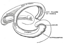
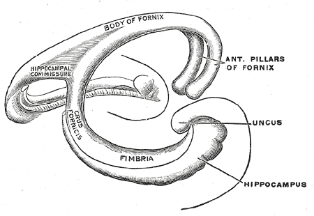
Diagram of the fornix. 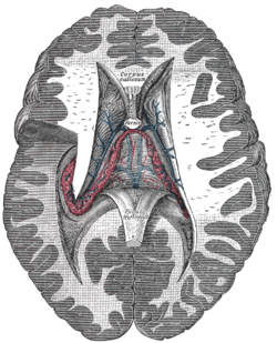
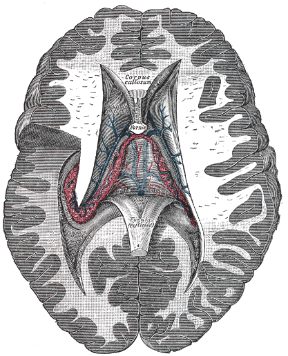
Tela chorioidea of the third ventricle, and the choroid plexus of the left lateral ventricle, exposed from above. Latin crus fornicis Gray's subject #189 838 NeuroNames hier-254 The crura (posterior pillars) of the fornix are prolonged backward from the body.
They are flattened bands, and, at their commencement, are intimately connected with the under surface of the corpus callosum.
Diverging from one another, each curves around the posterior end of the thalamus, and passes downward and forward into the inferior cornu of the lateral ventricle.
Here, it lies along the concavity of the hippocampus, on the surface of which some of its fibers are spread out to form the alveus, while the remainder are continued as a narrow white band, the fimbria hippocampi, which is prolonged into the uncus of the hippocampal gyrus.
This article was originally based on an entry from a public domain edition of Gray's Anatomy. As such, some of the information contained within it may be outdated.
Human brain, cerebrum, Interior of the cerebral hemispheres, white matter: commissural fibers and septum (TA A14.1.09.241–271, 569–571, GA 9.828, 838–840) Corpus callosum Lamina terminalis Fornix Septum pellucidum 
This anatomy article is a stub. You can help Wikipedia by expanding it.