- Median arcuate ligament
-
Median arcuate ligament 
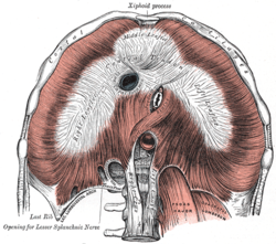
The diaphragm. Under surface. Latin ligamentum arcuatum medianum The median arcuate ligament is a ligament under the diaphragm that connects the right and left crura of diaphragm.
Contents
Structure
The median arcuate ligament is formed by the right and left crura of the diaphragm. The crura connect to form an arch, behind which is the aortic hiatus, through which pass the aorta, the azygos vein, and the thoracic duct.
Clinical significance
Compression of celiac artery and celiac ganglia by the median arcuate ligament can lead to the median arcuate ligament syndrome, which is characterized by abdominal pain, weight loss, and an epigastric bruit.[1]
See also
References
- ^ Duncan AA (April 2008). "Median arcuate ligament syndrome". Curr Treat Options Cardiovasc Med 10 (2): 112–6. doi:10.1007/s11936-008-0012-2. PMID 18325313. http://www.treatment-options.com/1092-8464/10/112.[dead link]
External links
- Median+arcuate+ligament at eMedicine Dictionary
- SUNY Figs 40:04-08 - "The abdominal surface of the diaphragm."
- 1738145849 at GPnotebook
- posteriorabdomen at The Anatomy Lesson by Wesley Norman (Georgetown University) (posteriorabdmus&nerves)
This article was originally based on an entry from a public domain edition of Gray's Anatomy. As such, some of the information contained within it may be outdated.
Thoracic diaphragm (TA A04.4.02, GA 4.404) General Openings major: Caval opening · Esophageal hiatus · Aortic hiatus
minor: Sternocostal triangle · Lumbocostal triangleCategories:- Ligaments of the torso
- Thoracic diaphragm
- Ligament stubs
Wikimedia Foundation. 2010.
