Posterior interventricular artery
- Posterior interventricular artery
Infobox Artery
Name = PAGENAME
Latin = ramus interventricularis posterior arteriae coronariae dextrae
GraySubject = 142
GrayPage = 547
Caption = Base and diaphragmatic surface of heart. (Posterior descending artery not visible, but it runs near the middle cardiac vein, which is labeled at the bottom.)
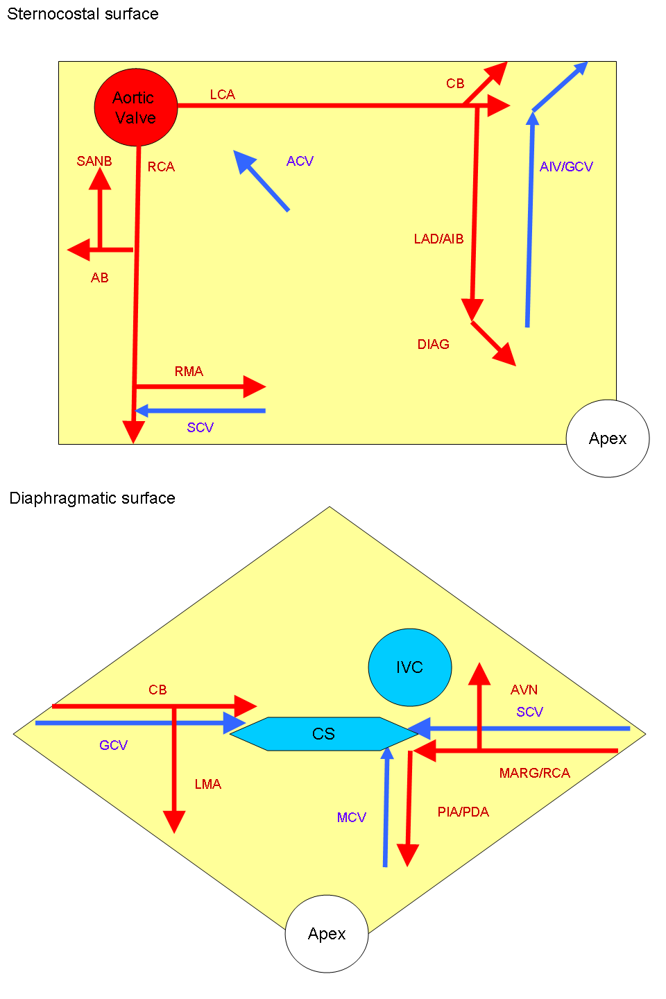
Caption2 = ARTERIES:
RCA = right coronary
AB = atrial branches
SANB = sinuatrial nodal
RMA = right marginal
LCA = left coronary
CB = circumflex branch
LAD/AIB = anterior interventricular
LMA = left marginal
PIA/PDA = posterior descending
AVN = atrioventricular nodal
VEINS:
SCV = small cardiac
ACV = anterior cardiac
AIV/GCV = great cardiac
MCV = middle cardiac
CS = coronary sinus
BranchFrom = right coronary artery
BranchTo =
Vein = middle cardiac vein
Supplies = ventricles
interventricular septum
System =
MeshName =
MeshNumber =
DorlandsPre = a_62
DorlandsSuf = 12157870
The posterior interventricular artery (PIV) (or posterior descending artery(PDA)) is typically a branch of the right coronary artery (80%, known as right dominance) which runs in the posterior interventricular sulcus to the apex of the heart where it meets with the anterior interventricular artery. Alternately, the PIV can be a branch of the circumflex coronary artery (20%, known as left dominance) which itself is a branch of the left coronary artery.External links
*
* - "Posterior view of the heart."
* [http://mywebpages.comcast.net/wnor/heartpostmajorvessels.jpgImage]
*
* [http://www.guidant.com/condition/heart/images/figure11_lg.jpgImage at guidant.com]
Wikimedia Foundation.
2010.
Look at other dictionaries:
Posterior interventricular sulcus — Infobox Anatomy Name = PAGENAME Latin = sulcus interventricularis anterior GraySubject = 138 GrayPage = 527 Caption = Base and diaphragmatic surface of heart. (Sulcus visible at center but not labeled.) Caption2 = System = Precursor = MeshName =… … Wikipedia
Interventricular septum — Section of the heart showing the ventricular septum. Interior of dorsal half … Wikipedia
Artery — A vessel that carries blood that is high in oxygen content away from the heart to the farthest reaches of the body. Since blood in arteries is usually full of oxygen, the hemoglobin in the red blood cells is oxygenated. The resultant form of… … Medical dictionary
Circumflex branch of left coronary artery — Artery: Circumflex branch of left coronary artery Base and diaphragmatic surface of heart. (Circumflex branch not visible, but would be near the coronary sinus.) … Wikipedia
Posterior pituitary — Pituitary gland. Posterior pituitary is in blue. Pars nervosa and infundibular stalk are not labeled, but pars nervosa is at bottom and infundibular stalk is at top.) … Wikipedia
Left marginal artery — Artery: Left marginal artery Base and diaphragmatic surface of heart. (Left marginal artery not visible, but would be near center left.) … Wikipedia
Right coronary artery — Artery: Right coronary artery Sternocostal surface of heart. (Right coronary artery visible at left.) Latin arteria coronaria dextra Gray s … Wikipedia
Deep cervical artery — Artery: Deep cervical artery Costocervical trunk with branches. Right side. Latin arteria cervicalis profunda Gray s … Wikipedia
Common carotid artery — Artery: Common carotid artery Schematic of the proximal aorta and its branches … Wikipedia
Right marginal branch of right coronary artery — Artery: Right marginal branch of right coronary artery Sternocostal surface of heart. (Right marginal artery not labeled, but is visible at bottom left.) … Wikipedia