- Molecular beacon
-
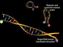 Structure of molecular beacons in their native conformations (top) or hybridized with a DNA strand (bottom)
Molecular beacons are oligonucleotide hybridization probes that can report the presence of specific nucleic acids in homogenous solutions. The terms more often used is molecular beacon probes. Molecular beacons are hairpin shaped molecules with an internally quenched fluorophore whose fluorescence is restored when they bind to a target nucleic acid sequence. This is a novel nonradioactive method for detecting specific sequences of nucleic acids. They are useful in situations where it is either not possible or desirable to isolate the probe-target hybrids from an excess of the hybridization probes.
Contents
Molecular beacon probes

A typical molecular beacon probe is 25 nucleotides long. The middle 15 nucleotides are complementary to the target DNA or RNA and do not base pair with one another, while the five nucleotides at each terminus are complementary to each other rather than to the target DNA. A typical molecular Beacon Structure can be divided in 4 parts:
- Loop: This is the 18–30 base pair region of the molecular beacon which is complementary to the target sequence.
- Stem: The beacon stem is formed by the attachment, to both termini of the loop, of two short (5 to 7 nucleotide residues) oligonucleotides that are complementary to each other.
- 5' fluorophore: At the 5' end of the molecular beacon, a fluorescent dye is covalently attached.
- 3' quencher (non fluorescent): The quencher dye is covalently attached to the 3' end of the molecular beacon. When the beacon is in closed loop shape, the quencher resides in proximity to the fluorophore, which results in quenching the fluorescent emission of the latter.
If the nucleic acid to be detected is complementary to the strand in the loop, the event of hybridization occurs. The duplex formed between the nucleic acid and the loop is more stable than that of the stem because the former duplex involves more base pairs. This causes the separation of the stem and hence of the fluorophore and the quencher. Once the fluorophore is distantiated from the quencher, illumination of the hybrid with light results in the fluorescent emission. The presence of the emission reports that the event of hybridization has occurred and hence the target nucleic acid sequence is present in the test sample.
Synthesis
Main article: Oligonucleotide synthesisMolecular beacons are synthetic oligonucleotides whose preparation is well documented. In addition to the conventional set of nucleoside phosphoramidites, the synthesis also requires a solid support derivatized with a quencher and a phosphoramidite building block designed for the attachment of a protected fluorescent dye.
Applications of molecular beacons
- SNP detection
- Real-time nucleic acid detection
- Real-time PCR quantification
- Allelic discrimination and identification
- Multiplex PCR assays
- Diagnostic clinical assays
References
Categories:- Biochemistry methods
- Fluorescence
Wikimedia Foundation. 2010.