- Fuligo septica
-
Fuligo septica Scientific classification Kingdom: Amoebozoa Phylum: Mycetozoa Class: Myxomycetes Order: Physarales Family: Physaraceae Genus: Fuligo Species: Fuligo septica
(L.) F.H.Wigg (1780)Synonyms[1] Mucor septicus L. (1763)
Reticularia septica (L.) With. (1792)
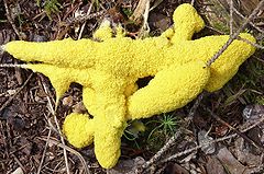
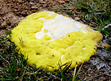
Aethalium septicum (L.) Fr. (1829)Fuligo septica is a species of plasmodial slime mold, and a member of the Myxomycetes class. It is commonly known as the dog vomit slime mold[2] or scrambled egg slime because of its peculiar yellowish, bile-colored appearance. A common species with a worldwide distribution, it is often found on bark mulch in urban areas after heavy rain or excessive watering. Their spores are produced on or in aerial sporangia and are spread by wind.
Contents
History and taxonomy
The first description of the species was provided by French botanist Jean Marchant in 1727, who referred to it as "flowers of tan"; Marchant classified it as a "sponge".[3] Carl Linnaeus called it Mucor septicus in his 1763 Species Plantarum.[4] The species was transferred to the genus Fuligo by German botanist Friedrich Heinrich Wiggers in 1780.[5]
Description and habitat
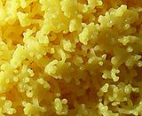
Like many slime molds, the cells of this species typically aggregate to form a plasmodium, a multinucleate mass of undifferentiated cells that may move in an ameboid-like fashion during the search for nutrients. F. septicas plasmodium may be anywhere from white to yellow-gray,[6] typically 2.5–20 cm (1.0–7.9 in) in diameter, and 1–3 cm (0.4–1.2 in) thick.[7] The plasmodium eventually transforms into a sponge-like aethalium, analogous to the spore-bearing fruiting body of a mushroom; which then degrades, darkening in color, and releases its dark-colored spores. This species is known to have its spores dispersed by beetles (family Lathridiidae).[8]
The spores have a two-layered wall, with a dense outer layer with spines, and a fibrous inner layer. During germination, the outer layer splits to create an opening, and more elastic inner layer ruptures later as protoplasm emerges. A remnant of the inner layer may be persistent and adhere to the protoplast after it has emerged from the spore. A peroxidase enzyme present in the inner cell wall plays a role in germination.[9]
Fuligo septica grows on rotten wood and plant debris, but can also grow on the leaves and stems of living plants.[10]
Human pathogenicity
This species is known to cause asthma and allergenic rhinitis in susceptible people.[11][12]
Resistance to metal toxicity
Slime molds have a high resistance to toxic levels of metals; one author was prompted to write "The levels of Zn in Fuligo septica were so high (4,000–20,000 ppm) that it is difficult to understand how a living organism can tolerate them."[13] The resistance to extreme levels of zinc appears to be unique to F. septica.[14] The mechanism of this metal resistance is now understood: F. septica produces a yellow pigment called fuligorubin A, which has been shown to chelate metals and convert them to inactive forms.[15]
Model of RNA processing
Introns are sections of DNA that must be properly cleaved, digested and processed prior to rendering functional mRNAs for protein synthesis. Because it has a large number of group I introns, F. septica is used as a model to understand the processing and evolution of RNA.[16][17]
Bioactive compounds
Extracts from F. septica show antibiotic activity against Bacillus subtilis and Candida albicans, and cytotoxic activity on KB cells (a cell line derived from a human carcinoma of the nasopharynx).[18]
Fuligo septica contains a yellow pigment called fuligorubin A that is thought to be involved in photoreception and in the process of energy conversion during its life cycle.[19] In 2011 a Japanese research group reported isolating and characterizing a new chlorine-containing yellow pigment from form flava that they called dehydrofuligoic acid.[20]
References
- ^ "Fuligo septica (L.) F.H. Wigg. 1780". MycoBank. International Mycological Association. http://www.mycobank.org/MycoTaxo.aspx?Link=T&Rec=149977. Retrieved 2011-03-25.
- ^ Armstrong M. (2007). Wildlife and Plants: Sea Urchin to Sponge. Marshall Cavendish. p. 998. ISBN 0761477101. http://books.google.com/books?id=jBh0jgWdWtsC&pg=PA998.
- ^ Ainsworth GC. (1976). Introduction to the History of Mycology. Cambridge, UK: Cambridge University Press. p. 60. ISBN 0-521-21013-5. http://books.google.com/books?id=3R09AAAAIAAJ&pg=PA60.
- ^ Linnaeus C. (1763). Species Plantarum (2 ed.). Stockholm: Impensis Direct. Laurentii Salvii. p. 1656. http://biodiversitylibrary.org/page/11835043.
- ^ Wiggers FH, Weber GH. (1780) (in Latin). Primitiae Florae Holsaticae. Litteris Mich. Frider. Bartschii Acad. Typogr.. p. 112. http://books.google.com/books?id=NNwXAAAAYAAJ&pg=PA112.
- ^ Kambly PE. (1939). "The color of myxomycete plasmodia". Botany 26 (6): 386–90.
- ^ Emberger G. (2008). "Fuligo septica". Fungi on Wood. http://www.messiah.edu/Oakes/fungi_on_wood/crust%20and%20parchment/species%20pages/Fuligo%20septica.htm. Retrieved 2008-12-08.
- ^ Blackwell M, Laman TG. (1982). "Spore dispersal of Fuligo septica (Myxomycetes) by Lathridiid beetles". Mycotaxon 14 (1): 58–60. http://www.cybertruffle.org.uk/cyberliber/59575/0014/001/0058.htm.
- ^ Stempen H, Evans RC. (1982). "Behavior of the inner wall layer of the germinating Fuligo septica spore: evidence of peroxidase activity". Mycologia 74 (1): 26–35. JSTOR 3792625. http://www.cybertruffle.org.uk/cyberliber/59350/0074/001/0026.htm.
- ^ Healy RA, Huffman DR., Tiffany LH, Knaphaus G. (2008). Mushrooms and Other Fungi of the Midcontinental United States (Bur Oak Guide). Iowa City, Iowa: University of Iowa Press. p. 340. ISBN 1-58729-627-6. http://books.google.com/books?id=tl2fVAHuej4C&pg=PA341.
- ^ Santili J, Rockwell WJ, Collins RP. (1895). "The significance of the spore of the Basidiomycetes (mushrooms and their allies) in bronchial asthma and allergenic rhinitis". Annals of Allergy 55: 469–71.
- ^ Gianini EH, Northy WT, Leathers CR. (1975). "The allergenic significance of certain fungi rarely reported as allergens". Annals of Allergy 35 (6): 372–6. PMID 1239229.
- ^ Setala A, Nuorteva P. (1989). "High metal contents found in Fuligo septica L. Wiggers and some other slime molds (Myxomycetes)". Karstenia 29 (1): 37–44.
- ^ Zhulidov DA, Robarts RD, Zhulidov AV, Zhulidova OV, Markelov DA, Rusanov VA, Headley JV. (2002). "Zinc accumulation by the slime mold Fuligo septica (L.) Wiggers in the former Soviet Union and North Korea". Journal of Environmental Quality 31 (3): 1038–42. doi:10.2134/jeq2002.1038. PMID 12026071. http://jeq.scijournals.org/cgi/pmidlookup?view=long&pmid=12026071.
- ^ Latowski D, Lesiak A, Jarosz-Krzeminska E, Strzalka K. (2008). "Fuligo septica, as a new model organism in studies on interaction between metal ions and living cells". Metal Ions in Biology and Medicine and Medicine 10: 204–9.
- ^ Lundblad EW, Einvik C, Rønning S, Haugli K, Johansen S. (2004). "Twelve Group I introns in the same pre-rRNA transcript of the myxomycete Fuligo septica: RNA processing and evolution". Molecular Biology and Evolution 21 (7): 1283–93. doi:10.1093/molbev/msh126. PMID 15034133. http://mbe.oxfordjournals.org/cgi/pmidlookup?view=long&pmid=15034133.
- ^ Haugen P, Coucheron DH, Rønning SB, Haugli K, Johansen S (2003). "The molecular evolution and structural organization of self-splicing group I introns at position 516 in nuclear SSU rDNA of myxomycetes". Journal of Eukaryotic Microbiology 50 (4): 283–92. doi:10.1111/j.1550-7408.2003.tb00135.x. PMID 15132172.
- ^ Pereira EC, Cavalcanti LDH, Campos-Takaki GMD, Nascimento, Silene CD. (1992). "Antibiotic and cytotoxic activities of crude extracts from Fuligo septica (L.) Wigg. and Tubifera microsperma (Berk. and Curt.) Martin (Myxomycetes)". Revista de Ciencias Biomedicas (13): 23–32.
- ^ Rahman A. (1988). Studies in Natural Products Chemistry. Amsterdam: Elsevier. pp. 237–8. ISBN 0-444-51510-0.
- ^ Shintani A, Toume K, Yamamoto Y, Ishibashi M. (2010). "Dehydrofuligoic acid, a new yellow pigment isolated from the myxomycete Fuligo septica f. flava". Heterocycles 82 (1): 839–42. ISSN 0385-5414.
External links
Categories:
Wikimedia Foundation. 2010.


