Optic vesicles

Optic vesicles

Infobox Embryology
Name = PAGENAME
Latin = v. ophthalmica
GraySubject = 224
GrayPage = 1001


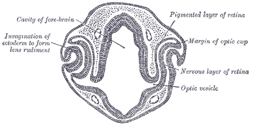
Caption = Transverse section of head of chick embryo of forty-eight hours’ incubation. (Optic vesicle labeled at lower right.)


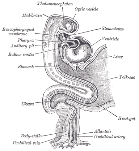
Caption2 = Human embryo about fifteen days old. Brain and heart represented from right side. Digestive tube and yolk sac in median section. (Optic vesicle labeled at center top.)
System =
CarnegieStage = 11
Precursor =
GivesRiseTo =
MeshName =
MeshNumber =
DorlandsPre = v_07
DorlandsSuf = 12855707
The eyes begin to develop as a pair of diverticula from the lateral aspects of the forebrain. These diverticula make their appearance before the closure of the anterior end of the neural tube; after the closure of the tube they are known as the optic vesicles.

They project toward the sides of the head, and the peripheral part of each expands to form a hollow bulb, while the proximal part remains narrow and constitutes the optic stalk.


=Additional

External links

*
* [http://www.vision.ca/eye/o.cup.l.vesicle.html Overview at vision.ca]
* [http://isc.temple.edu/neuroanatomy/lab/embryo/eye2.htm Overview at temple.edu]


Wikimedia Foundation. 2010.

Игры ⚽ Нужна курсовая?

Look at other dictionaries:

  • Optic cup (embryology) — Transverse section of head of chick embryo of forty eight hours’ incubation. (Margin of optic cup labeled at upper right.) …   Wikipedia

  • Optic stalk — Transverse section of head of chick embryo of fifty two hours’ incubation …   Wikipedia

  • Optic vesicle — Transverse section of head of chick embryo of forty eight hours’ incubation. (Optic vesicle labeled at lower right.) …   Wikipedia

  • animal development — Introduction  the processes that lead eventually to the formation of a new animal starting from cells derived from one or more parent individuals. Development thus occurs following the process by which a new generation of organisms is produced by …   Universalium

  • Eye development — The eye develops from the neural tube, the epidermis, and the periocular mesenchyme, which receives contributions from both the neural crest and mesoderm lineages. equential inductionsThis development is an example of sequential inductions where… …   Wikipedia

  • Collie eye anomaly — (CEA) is a congenital, inherited, bilateral eye disease of dogs involving the retina, choroid, and sclera. It can be a mild disease or cause blindness. CEA is caused by a simple autosomal recessive gene defect. There is no treatment. Contents 1… …   Wikipedia

  • Neural tube — Transverse section of half of a chick embryo of forty five hours incubation. The dorsal (back) surface of the embryo is towards the top of this page, while the ventral (front) surface is towards the bottom. (Neural tube is in green.) …   Wikipedia

  • Cephalopod — Cephalopods Temporal range: Devonian – Recent[1] (Stem groups from Cambrian)[2] …   Wikipedia

  • Vesicle — may refer to:* Synaptic vesicle * Auditory vesicle * Optic vesicles * Seminal vesicle * Subsporangial vesicle * Vesical arteries * Vesicle (biology), a relatively small and enclosed compartment within a cell * Vesicle (dermatology), a liquid… …   Wikipedia

  • di|en|ceph|a|lon — «DY ehn SEHF uh lon», noun. Anatomy. the posterior part of the forebrain, connecting the cerebrum to the midbrain and including the optic vesicles and pineal body; interbrain. ╂[< New Latin diencephalon < Greek diá through + enképhalon… …   Useful english dictionary

Share the article and excerpts

Direct link
Do a right-click on the link above
and select “Copy Link”