- Movat's stain
-
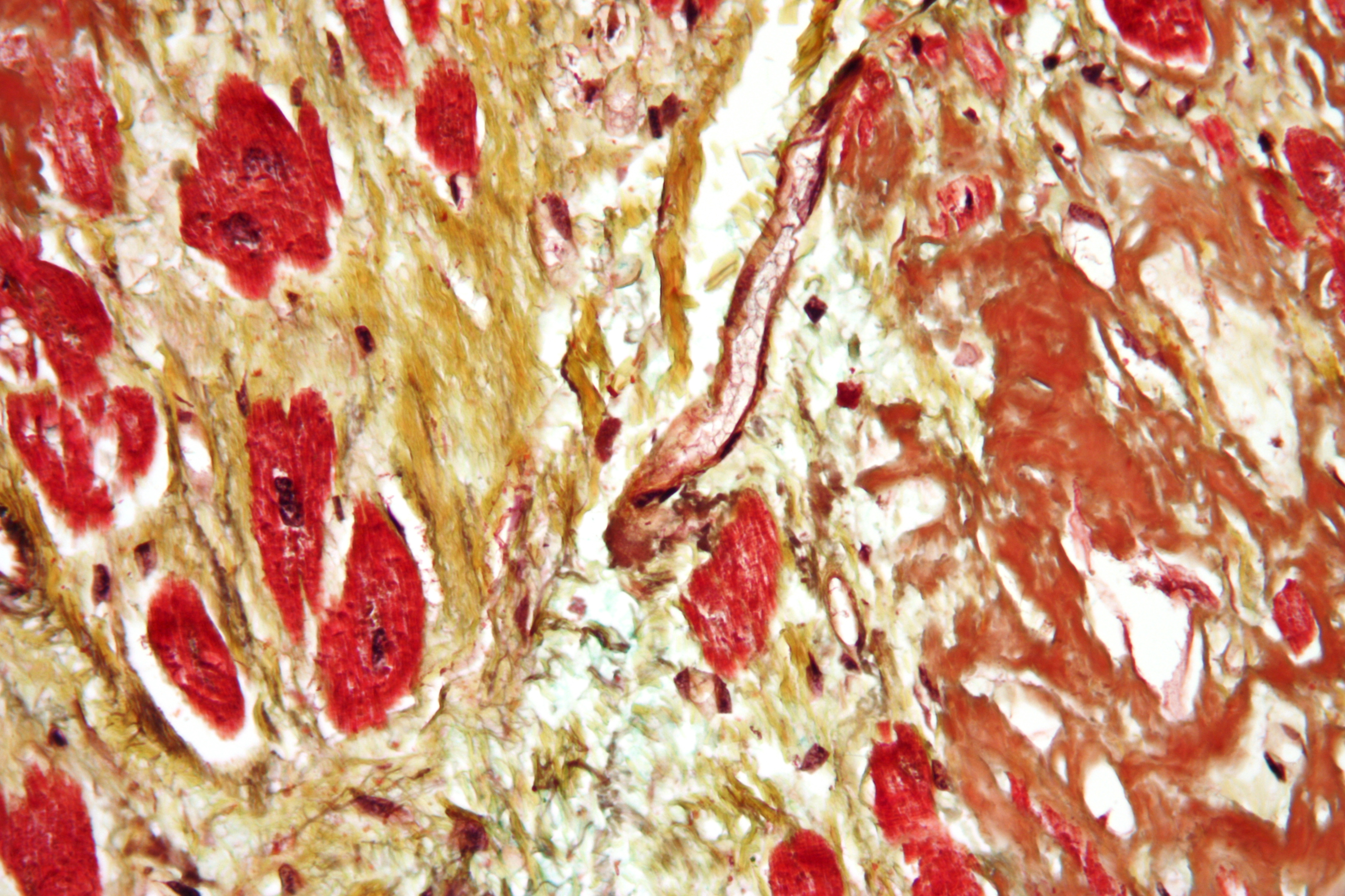
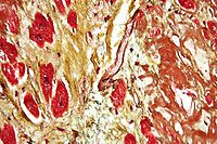 Movat's stain showing amyloid (brown) and fibrosis (yellow) of the heart.
In pathology, the Movat's stain is a staining method in histology that is based on Alcian blue.[1] It is used in cardiovascular pathology.
Contents
Interpretation
- Black = nuclei, elastic fibres
- Yellow = collagen, reticular fibers
- Blue = ground substance, mucin
- Bright red = fibrin
- Red = muscle
Additional images
-
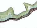
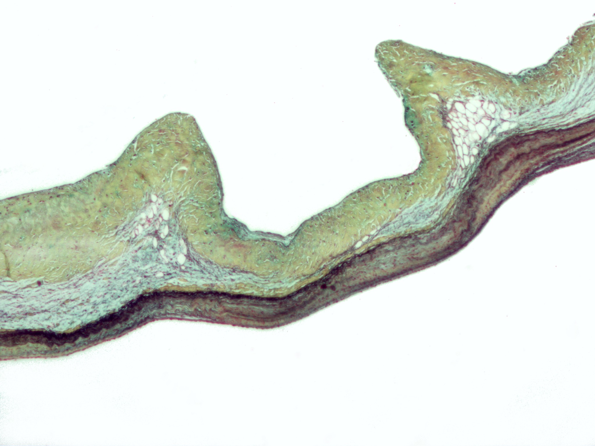
Movat's stain demonstrating thickening of the spongiosa layer (blue) in myxomatous degeneration of the aortic valve.
References
- ^ Modified Movat's Pentachrome Stain. University of Pennsylvania. URL: http://www.med.upenn.edu/mcrc/histology_core/movat.shtml. Accessed on: 4 December 2009.
See also
Categories:- Pathology stubs
- Staining
Wikimedia Foundation. 2010.