- Microtia
-
For the orchid genus, see Microtis.
Microtia Classification and external resources 
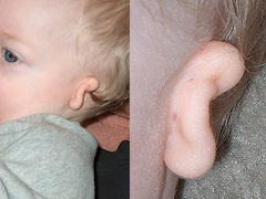
Unilateral grade III microtia (more often affecting the right ear).ICD-10 Q17.2 ICD-9 744.23 OMIM 600674 DiseasesDB 29876 eMedicine ped/3003 Microtia, also called microtia-anotia,[1] is a congenital deformity where the pinna (outer ear) is very small and underdeveloped (microtia) or absent (anotia). It can be unilateral (one side only) or bilateral (affecting both sides). It occurs in 1 out of about 8,000–10,000 births. In unilateral microtia, the right ear is most commonly affected. It can occur as a complication of taking Accutane (isotretinoin) during pregnancy.[2]
Contents
Classification
There are four grades of microtia:[3]
- Grade I: A slightly small ear with identifiable structures and a small but present external ear canal
- Grade II: A partial or hemi-ear with a closed off or stenotic external ear canal producing a conductive hearing loss
- Grade III: Absence of the external ear with a small peanut vestige structure and an absence of the external ear canal and ear drum
- Grade IV: Absence of the total ear or anotia.
Treatment
Grade III microtia is most common,[4] and can be corrected by surgery. Typically, testing is first done to determine the quality of hearing. This can be done as early as in the first two weeks with a BAER test (Brain Stem Auditory Response Test).[5][6] At age 5-6, CT or "CAT Scans" of the middle ear can be done to elucidate its development and clarify which patients are appropriate candidates for surgery to improve hearing. For younger individuals, this is done under sedation. The age when outer ear surgery can be attempted depends upon the technique chosen. The earliest is 7 for Rib Cartilage Grafts. However, some surgeons recommend waiting until a later age, such as 8-10 when the ear is closer to adult size.
Surgical options
There are two separate issues regarding the ear:
- Auricular reconstruction to restore the visual appearance and form of the outer ear
- Repair of atresia or application of a Bone Conduction Hearing Solution (BAHA) to restore hearing.
The hearing loss associated with congenital aural atresia is a conductive hearing loss—hearing loss caused by inefficient conduction of sound to the inner ear. Essentially, children with aural atresia have hearing loss because the sound cannot travel into the (usually) healthy inner ear—there is no ear canal, no eardrum, and the small ear bones (malleus/hammer, incus/anvil, and stapes/stirrup) are underdeveloped. "Usually" is in parentheses because rarely, a child with atresia also has a malformation of the inner ear leading to a sensorineural hearing loss (as many as 19% in one study).[7] Sensorineural hearing loss is caused by a problem in the inner ear, the cochlea. Sensorineural hearing loss is not correctable by surgery, but properly fitted and adjusted hearing amplification (hearing aids) generally provide excellent rehabilitation for this hearing loss. If the hearing loss is severe to profound in both ears, the child may be a candidate for a cochlear implant (beyond the scope of this discussion).
Unilateral sensorineural hearing loss was not generally considered a serious disability by the medical establishment before the nineties; it was thought that the afflicted person was able to adjust to it from birth. In general, there are exceptional advantages to gain from an intervention to enable hearing in the microtic ear, especially in bilateral microtia. Children with untreated unilateral sensorineural hearing loss are more likely to have to repeat a grade in school and/or need supplemental services (e.g., FM system - see below) than their peers.[8][9]
Children with unilateral sensorineural hearing loss often require years of speech therapy in order to learn how to enunciate and understand spoken language. What is truly unclear, and the subject of an ongoing research study, is the effect of unilateral conductive hearing loss (in children with unilateral aural atresia) on scholastic performance. If atresia surgery or some form of amplification is not used, special steps should be taken to ensure that the child is accessing and understanding all of the verbal information presented in school settings. Recommendations for improving a child's hearing in the academic setting include preferential seating in class, an FM system (the teacher wears a microphone, and the sound is transmitted to a speaker at the child's desk or to an ear bud or hearing aid the child wears), a bone conducting hearing aid, or conventional hearing aids. Age for BAHA implantation depends on whether you are in Europe (18 months) or the US (age 5). Until then it is possible to fit a Baha on a softband[10][11]
It is important to note that not all children with aural atresia are candidates for atresia repair. Candidacy for atresia surgery is based on the hearing test (audiogram) and CT scan imaging.
In cases where a later surgical reconstruction of the external ear of the child might be possible, positioning of the Baha implant is critical. It may be necessary to position the implant further back than usual to enable successful reconstructive surgery – but not so far as to compromise hearing performance. If the reconstruction is ultimately successful, it is easy to remove the percutaneous Baha abutment. If the surgery is unsuccessful, the abutment can be replaced and the implant re-activated to restore hearing.
For auricular reconstruction, there are several different options:
- Rib Cartilage Graft Reconstruction: This surgery may be performed by specialists in the technique.[12][13][14][15][16] It involves sculpting the patient's own rib cartilage into the form of an ear. Because the cartilage is the patient's own living tissue, the reconstructed ear continues to grow as the child does. In order to be sure that the rib cage is large enough to provide the necessary donor tissue, some surgeons wait until the patient is 8 years of age.;[15][16] however, some surgeons with more experience with this technique may begin the surgery on a child aged six.[13][14][17] This surgery varies from two to four stages depending on the surgeon's preferred method. The major advantage of this surgery is that the patient's own tissue is used for the reconstruction.
- Reconstruct the ear using a polyethylene plastic implant (also called Medpor): This is a 1-2 stage surgery that can start at age 3 and can be done as an outpatient without hospitalization. Using the porous framework which allows the patient's tissue to grow into the material and the patient's own tissue flap, a new ear is constructed in a single surgery. A small second surgery is performed in 3–6 months if needed for minor adjustments. This surgery should only be performed by experts in the techniques involved.[18]
- Ear Prosthesis: This surgery is ideally suited to those in whom other techniques have failed or are severely scarred by traumatic injuries such as burns.[4] An auricular (ear) prosthesis is custom made by an anaplastologist to mirror the other ear.[19] Prosthetic ears can appear very realistic. They require a few minutes of daily care. They are typically made of silicone, which is colored to match the patient's skin and can be attached using either adhesive or with titanium screws inserted into the skull to which the prosthetic is attached with a magnetic or bar/clip type system. These screws are the same as the BAHA (Bone Anchored Hearing Aid) screws and can be placed simultaneously.
Related Conditions
Aural atresia is the underdevelopment of the middle ear and canal and usually occurs in conjunction with microtia. Atresia occurs because patients with microtia may not have an external opening to the ear canal, though. However, the cochlea and other inner ear structures are usually present. The grade of microtia usually correlates to the degree of development of the middle ear.[6] [20] Microtia is usually isolated, but may occur in conjunction with hemifacial microsomia, Goldenhar Syndrome or Treacher-Collins Syndrome.[21]
Microtia can cause difficulties with wearing headphones and glasses.[22] It is also occasionally associated with kidney abnormalities (rarely life-threatening), and jaw problems, and more rarely, heart defects and vertebral deformities.[14]
If a canal is built where one does not exist, minor complications can arise from the body's natural tendency to heal an open wound closed. Repairing aural atresia is a very detailed and complicated surgical procedure which requires an expert in atresia repair.[20] While complications from this surgery can arise, the risk of complications is greatly reduced when using a highly experienced otologist. Atresia patients who opt for surgery will temporarily have the canal packed with gelatin sponge and silicone sheeting to prevent closure. It must be stressed that many surgeons believe that ear canal reconstruction is unnecessary and overcomplicated and that very good hearing is possible with modern hearing aids which can be hidden under the skin.
There are several organizations which provide information and support to affected people.
In some countries, the outer ear reconstruction is considered as cosmetic surgery, which means that it is not necessary, and hence is not paid for by health insurance.
References
- ^ Online 'Mendelian Inheritance in Man' (OMIM) Microtia-Anotia -600674
- ^ Pretest self assessment and review for the USMLE, pediatrics, 12th edition, question 84, general pediatrics
- ^ "What is microtia?". Patient Education & Resources: Health Library. New York Eye and Ear Infirmary. http://www.nyee.edu/faqlist.html?tablename=faq&key=60.
- ^ a b Brent B. (1992). "Auricular Repair with Autogenous Rib Cartilage Grafts: Two Decades of Experience with 600 Cases". Plastic & Reconstructive Surgery (90): 355.
- ^ De la Cruz A, Kesser BW. (1999). "Management of the Unilateral Atretic Ear". In Controversies in Otolaryngology—Head and Neck Surgery. Pensak M (ed.) New York: Thieme Medical Publishers. pp. 381–385.
- ^ a b Kountakis SE, Helidonis E, Jahrsdoerfer RA (August 1995). "Microtia grade as an indicator of middle ear development in aural atresia". Arch Otolaryngol Head Neck Surg 121 (8): 885–6. PMID 7619415.
- ^ Vrabec JT, Lin JW. (2010). "Inner Ear Anomalies in Congenital Aural Atresia". Otology/Neurotology 31: 1421.
- ^ Bess FH, Tharpe AM. (1986). "Case History Data on Unilaterally Hearing-Impaired Children". Ear and Hearing 7: 14.
- ^ Bess FH, Tharpe AM. (1988). "Performance and Management of Children with Unilateral Sensorineural Hearing Loss". Cand Audiol Supple 30: 75.
- ^ Nicholson N, Christensen L, Dornhoffer J, Martin P, Smith-Olinde L. Verification of Speech Spectrum Audibility for Pediatric Baha Softband Users with Craniofacial Anomalies. Cleft Palate Craniofacial Journal. 2010 Feb;22.
- ^ Verhagen CV, Hol MK, Coppens-Schellekens W, Snik AF, Cremers CW. The Baha Softband A new treatment for young children with bilateral congenital aural atresia. International Journal of Pediatric Otorhinolaryngology. 2008;72, 1455—1459.
- ^ Tanzer RC. (1959). "Total Reconstruction of the External Ear". Plastic & Reconstructive Surgery 23: 1.
- ^ a b Brent B. (1999). "Technical Advances with Autogenous Rib Cartilage Grafts—A Personal Review of 1,200 Cases". Plastic & Reconstructive Surgery 104: 319.
- ^ a b c Brent B. (1992). "Auricular Repair with Autogenous Rib Cartilage Grafts: Two Decades of Experience with 600 Cases". Plastic & Reconstructive Surgery 90: 355.
- ^ a b Firmin F. (1992). "Microtie Reconstruction par la Technique de Brent". Annals Chirurgie Plastica Esthetica 1: 119.
- ^ a b Nagata S. (1994). "Reconstruction of the Auricle". Plastic & Reconstructive Surgery 93: 225.
- ^ Brent B. (2000). "The Team Approach to Treating the Microtia-Atresia Patient". Otolarng (Clinics of North America) 33: 1353.
- ^ Reinisch JF, Lewin S (August 2009). "Ear reconstruction using a porous polyethylene framework and temporoparietal fascia flap". Facial Plast Surg 25 (3): 181–9. doi:10.1055/s-0029-1239448. PMID 19809950. http://www.thieme-connect.com/DOI/DOI?10.1055/s-0029-1239448.
- ^ Tanner PB, Mobley SR. (May 2006). "External Auricular and Facial Prosthetics: A Collaborative Effort of the Reconstructive Surgeon and Anaplastologist. Auricular Surgery: Aesthetic and Reconstructive". Facial Plast Surg Clin North Am 14(2): 137–45, vi–vii.
- ^ a b Jahrsdoerfer RA, Kesser BW. (1995). "Issues on Aural Atresia for the facial Plastic Surgeon". Facial Plastic Surgery (11): 274.
- ^ Huston Katsanis S, Cutting GR (July 2004). "Treacher Collins Syndrome". GeneReviews. PMID 20301704.
- ^ http://www.optometrists.asn.au/gui/files/ceo856389.pdf
External links
- Craniofacial Anomalies Program at Children's Hospital Boston
- Microtia Repair
- Atresia Repair — University of Virginia
- Microtia Reconstruction at Cedars-Sinai
- Bennun RD, Mulliken JB, Kaban LB, Murray JE (December 1985). "Microtia: a microform of hemifacial microsomia". Plast. Reconstr. Surg. 76 (6): 859–65. doi:10.1097/00006534-198512000-00010. PMID 4070453.
- Annual Atresia-Microtia Conference, October 11–12, 2009
- Goldenhar Family Support Group (UK)
- Microtia Australia
Congenital malformations and deformations of ears (Q16–Q17, 744.0–744.3) Size Position Other Accessory auricleM: EAR
anat(e/p)/phys/devp
noco/cong, epon
proc, drug(S2)
Categories:- Diseases of the ear and mastoid process
- Congenital disorders of eye, ear, face and neck
Wikimedia Foundation. 2010.