- Prevertebral fascia
Infobox Anatomy
Name = PAGENAME
Latin = lamina prevertebralis fasciae cervicalis
GraySubject = 111
GrayPage = 389
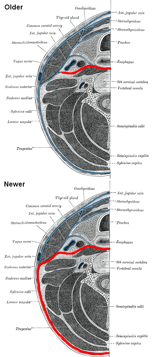
Caption = Prevertebral fascia labeled in red, both according to older litterature (e.g. Gray's) and newer litteratureEssential Clinical Anatomy. K.L. Moore & A.M. Agur. Lippincott, 2 ed. 2002. ] . Section of the neck at about the level of the sixth cervical vertebra. Showing the arrangement of the fascia coli.
Caption2 =
Precursor =
System =
Artery =
Vein =
Nerve =
Lymph =
MeshName =
MeshNumber =
DorlandsPre = l_02
DorlandsSuf = 12476606
The prevertebral fascia (or prevertebral layer of cervical fascia) is afascia in theneck .Variations
In some literature, the prevertebral fascia also includes the rest of the fascia extending around the
vertebral column and enclosing all muscles laterally and posteriorly to itEssential Clinical Anatomy. K.L. Moore & A.M. Agur. Lippincott, 2 ed. 2002. ] . However, in this article, it is assumed to be as marked in picture.Location
The prevertebral fascia extends medialward behind the carotid vessels, where it assists in forming their sheath, and passes in front of the
prevertebral muscles .The prevertebral fascia is fixed above to the
base of the skull , and below it extends behind the esophagus into theposterior mediastinal cavity of thethorax . It descends in front of thelongus colli muscles .The prevertebral fascia is prolonged downward and laterally behind the carotid vessels and in front of the
scaleni , and forms a sheath for thebrachial nerves andsubclavian vessels in the posterior triangle of the neck; it is continued under the clavicle as the axillary sheath and is attached to the deep surface of thecoracoclavicular fascia .urrounding structures
It forms the posterior limit of a fibrous compartment, which contains the
larynx and trachea, thethyroid gland , and thepharynx andesophagus .Parallel to the carotid sheath and along its medial aspect the prevertebral fascia gives off a thin lamina, the
buccopharyngeal fascia , which closely invests theconstrictor muscles of the pharynx , and is continued forward from theconstrictor pharyngis superior on to thebuccinator . This is attached to the prevertebral layer by loose connective tissue only, and thus an easily distended space, theretropharyngeal space , is found between them.Immediately above and behind the clavicle an areolar space exists between the investing layer and the sheath of the subclavian vessels, and in this space are found the lower part of the external jugular vein, the descending clavicular nerves, the transverse scapular and transverse cervical vessels, and the inferior belly of the Omohyoideus muscle.
This space is limited below by the fusion of the coracoclavicular fascia with the anterior wall of the
axillary sheath .References
External links
*
Wikimedia Foundation. 2010.