- Costodiaphragmatic recess
-
Costodiaphragmatic recess 
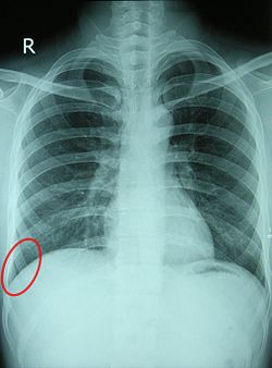
Chest X-ray of a 30-year-old healthy man, with the costodiaphragmatic recess label in red ellipse. 
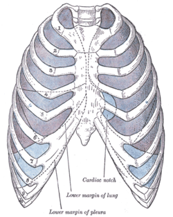
Front view of thorax, showing the relations of the pleuræ and lungs to the chest wall. Pleura in blue; lungs in purple. Latin recessus costodiaphragmaticus In the pleural cavity, the costodiaphragmatic recess (also called phrenicocostal sinus[1]) is a potential space at the posteriormost tips of the cavity, located at the junction of the costal pleura and diaphragmatic pleura. It measures approximately 5 cm vertically, and extends from the eighth to the tenth rib along the mid-axillary line.
The lungs expand into this recess during forced inspiration, however the recess never fills completely. During expiration, it contains no lung tissue, only pleural fluid.
Pleural effusions collect in the costodiaphragmatic recess when in standing position.[2]
A thoracocentesis (pleural tap) is often performed here while a patient is in full expiration because of less risk of puncturing the lungs and thereby causing pneumothorax.[2]
See also
References
- ^ drugs.com > phrenicocostal-sinus Retrieved May 2011
- ^ a b drugs.com > costodiaphragmatic-recess Retrieved May 2011
External links
- costodiaphragmatic+recess at eMedicine Dictionary
- 33947706 at GPnotebook
- Roche Lexicon - illustrated navigator, at Elsevier 02101.002-1
- Diagram (Question #4, item E)
This article was originally based on an entry from a public domain edition of Gray's Anatomy. As such, some of the information contained within it may be outdated.
Anatomy, Respiratory system: Thoracic cavity (TA A07, TH H3.05.03, GA 11.1087) Pleurae Parietal pleura (Cervical, Costal, Mediastinal, Diaphragmatic) · Visceral pleura · Pulmonary ligament · recesses (Costomediastinal, Costodiaphragmatic) · Pleural cavity
Suprapleural membraneMediastinum General Categories:- Respiratory system stubs
- Pleura
Wikimedia Foundation. 2010.
