- Oxidase test
-
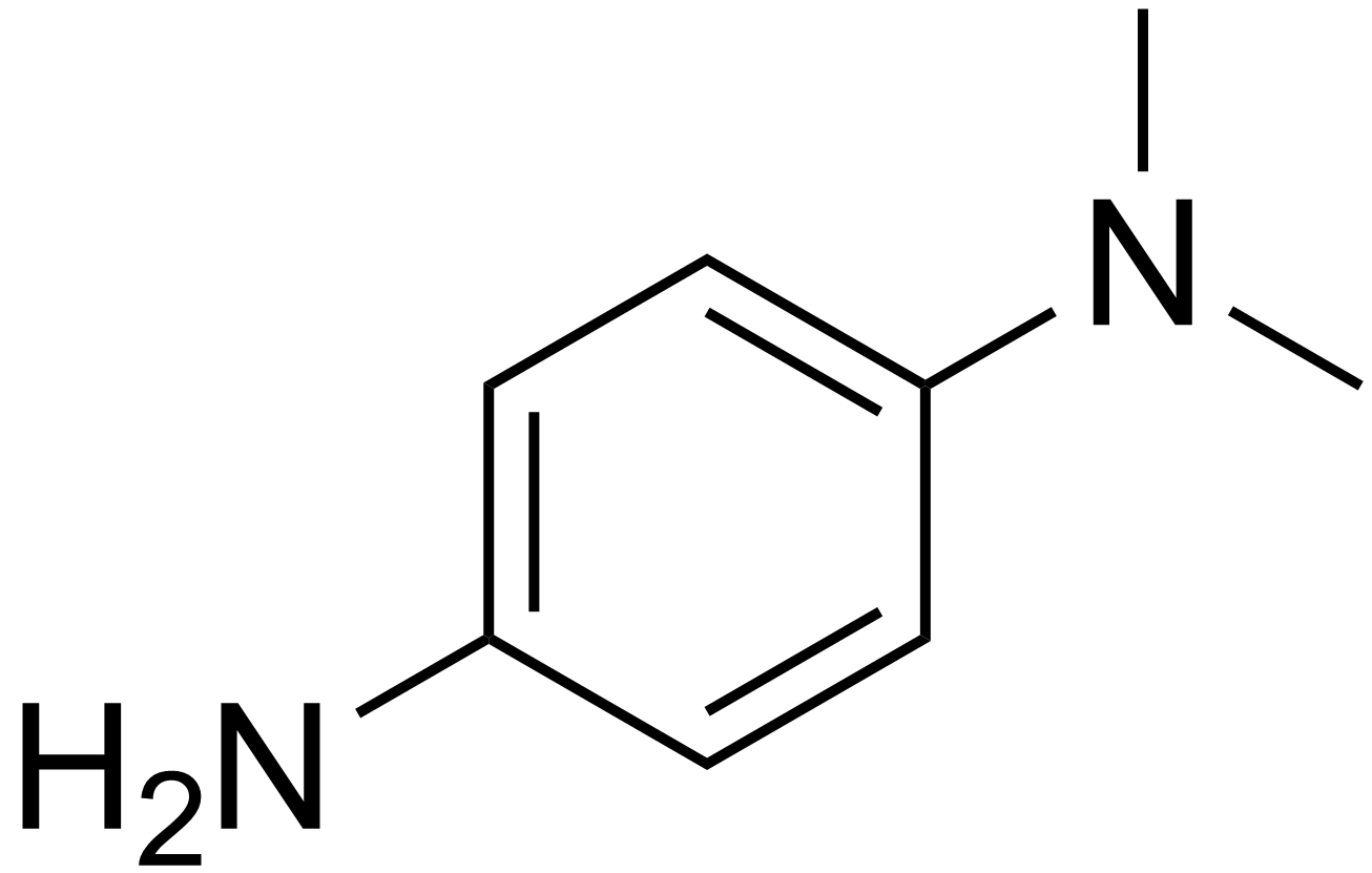
 DMPD
DMPD
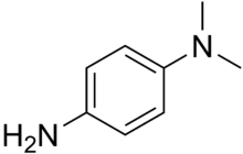
 TMPD
TMPDThe oxidase test is a test used in microbiology to determine if a bacterium produces certain cytochrome c oxidases.[1] It uses disks impregnated with a reagent such as N,N,N′,N′-tetramethyl-p-phenylenediamine (TMPD) or N,N-Dimethyl-p-phenylenediamine (DMPD), which is also a redox indicator. The reagent is a dark blue to maroon color when oxidized, and colorless when reduced.Oxidase positive bacteria possess cytochrome oxidase or indophenol oxidase (an iron containing hemoprotein)[2]. These both catalyze the transport of electrons from donor compounds (NADH) to electron acceptors (usually oxygen). The test reagent, N, N, N’, N’-tetra-methyl-p-phenylenediamine dihydrochloride acts as an artificial electron donor for the enzyme oxidase. The oxidized reagent forms the colored compound indophenol blue. The cytochrome system is usually only present in aerobic organisms which are capable of utilizing oxygen as the final hydrogen receptor. The end product of this metabolism is either water or hydrogen peroxide (broken down by catalase)[3].
Contents
Classification
Strains may either be oxidase positive (OX+) or negative (OX-).
OX+
OX+ normally means that the bacterium contains cytochrome c oxidase and can therefore utilize oxygen for energy production with an electron transfer chain.
Typically the Pseudomonadaceae are OX+[citation needed]
Another example is the preliminary identification of Neisseria and Moraxella genera, which are both oxidase positive, Gram-negative diplococci.
Many Gram-negative spiral curved rods are also oxidase positive, which includes Helicobacter pylori, Vibrio cholera, and Campylobacter jejuni.
Also Legionella pneumophila is oxidase positive. A trick to remember the most medical relevant bacteria is: "VIce President CHeNEy MOstly LEads" (Vibrio, Pseudomonas, Campylobacter, Helicobacter, Neisseria, Moraxella, and Legionella, respectively).
OX-
OX- normally means that the bacterium does not contain cytochrome c oxidase and therefore cannot utilize oxygen for energy production with an electron transfer chain.
Typically Enterobacteriaceae are OX-.[4]
Procedures
- Wet each disk with about 4 inoculating loops of de-ionized water.
- Use a loop to aseptically transfer a large mass of pure bacteria to the disk.
- Observe the disk for up to 3 minutes. If the area of inoculation turns dark blue to maroon to almost black, then the result is positive. If a color change does not occur within three minutes, the result is negative.
Alternatively, live bacteria cultivated on trypticase soy agar plates may be prepared using sterile technique with a single-line streak inoculation. The inoculated plates are incubated at 37°C for 24–48 hours to establish colonies. Fresh bacterial preparations should be used. After colonies have grown on the media, two-to-three drops of the reagent DMPD is added to the surface of each organism to be tested.
- A positive test (OX+) will result in a color change to pink, through maroon and into black, within 10–30 seconds.
- A negative test (OX-) will result in a light pink coloration or absence of coloration.
References
- ^ "Oxidase Test and Modified Oxidase Test". http://web.mst.edu/~microbio/Lab_Supplement/Oxidase.html. Retrieved 2008-11-07.
- ^ . Isenberg HD, editor. Clinical Microbiology Procedures Handbook. American Society for Microbiology; 2004. p. 3.3.2-3.3.2.13
- ^ MacFaddin JF, editor. Biochemical Tests for Identification of Medical Bacteria. 3rd ed. Philadelphia:Lippincott Williams and Wilkins; 2000. p. 363-7
- ^ Farmer JJ, Fanning GR, Huntley-Carter GP, et al. (May 1981). "Kluyvera, a new (redefined) genus in the family Enterobacteriaceae: identification of Kluyvera ascorbata sp. nov. and Kluyvera cryocrescens sp. nov. in clinical specimens". J. Clin. Microbiol. 13 (5): 919–33. PMC 273917. PMID 7240403. http://jcm.asm.org/cgi/pmidlookup?view=long&pmid=7240403.
External links
- Oxidase test video
- Oxidase Test Procedure
Categories:- Microbiology techniques
- Bacteria stubs

Wikimedia Foundation. 2010.